Plastination techniques: from three-dimensional specimens to transparent anatomical slices

Plastination is not a single preservation procedure, but rather a family of techniques based on a common principle: the replacement of water and part of the lipids within biological tissues by a curable polymer. Once this polymer has been introduced into the tissues and subsequently hardened, the resulting anatomical specimens are dry, odourless, durable, and suitable for long-term handling, teaching, research, or exhibition [1,2].

Although different plastination protocols and polymers have been developed since the technique was introduced by Gunther von Hagens in the late 1970s, three major approaches have become particularly important: silicone, epoxy, and polyester plastination. Each produces specimens with different physical and optical properties and, consequently, each is particularly suited to specific anatomical applications.

The Common Principles of Plastination

Regardless of the polymer used, conventional plastination follows a series of fundamental steps. First, the specimen is prepared and, in most cases, fixed to preserve its anatomical structures. The tissue is then dehydrated, usually with acetone, removing the water contained within it and part of its lipids. The central step of the process is forced impregnation. Under vacuum, the acetone within the specimen progressively vaporises and is replaced by a liquid polymer. The impregnated specimen is finally cured or hardened, transforming the polymer into a stable material permanently incorporated within the tissues [1]. The choice of polymer is crucial because it determines many of the final characteristics of the plastinated specimen, including its flexibility, transparency, mechanical resistance, and suitability for either three-dimensional preparations or anatomical slices.

Silicone Plastination: Preserving Anatomy in Three Dimensions

Silicone plastination is the most widely used plastination technique and is particularly suitable for preserving whole organs, body parts, dissected anatomical regions, and other three-dimensional specimens [1–3]. After forced impregnation with silicone, the specimen is positioned appropriately and the polymer is cured. The resulting preparations are generally opaque or only slightly translucent and retain a relatively natural appearance. Depending on the protocol and the tissues involved, they may also preserve a certain degree of flexibility.

One of the main advantages of silicone plastination is that it maintains the three-dimensional relationships between anatomical structures while producing specimens that can be handled repeatedly without the need for immersion in preservative fluids. This makes silicone plastinates especially useful for gross anatomy teaching, where students need to identify structures, follow vessels and nerves, understand anatomical relationships, and examine a specimen from different perspectives. For this reason, silicone plastination has become particularly valuable for the long-term preservation of carefully dissected specimens. A complex anatomical preparation that may require many hours of dissection can remain available for successive generations of students and can be used in practical classes, examinations, demonstrations, and exhibitions [2,3].
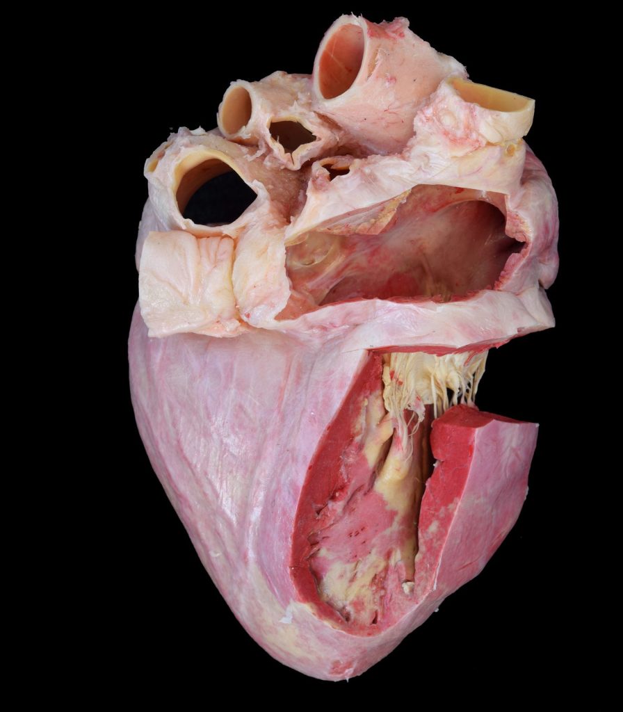
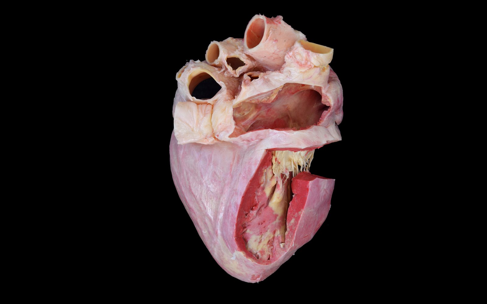
Pig heart preserved by silicone plastination, with its internal cavities exposed.
Figure 1.1. Pig heart preserved by silicone plastination, with its internal cavities exposed.

Epoxy Plastination: Anatomy in Transparent Sections

Epoxy plastination has a very different purpose. Rather than preserving complete three-dimensional preparations, epoxy resins are mainly used to produce thin, transparent anatomical slices. In epoxy sheet plastination, frozen anatomical material is sectioned into thin slices, commonly only a few millimetres thick. Following dehydration and forced impregnation with epoxy resin, the sections are cast and cured to produce firm, transparent preparations with a high degree of anatomical detail [1,4]. Their transparency is one of their most important characteristics. Structures can be examined within their original topographical context without destroying the relationships between tissues. Depending on section thickness details ranging from gross anatomical structures to considerably thinner morphological features can be investigated [4].

Epoxy plastination is therefore especially valuable for sectional and topographic anatomy and for correlating physical anatomical sections with diagnostic imaging techniques such as computed tomography (CT) and magnetic resonance imaging (MRI). The technique has consequently become an important tool not only for teaching but also for anatomical and morphological research [4,5].
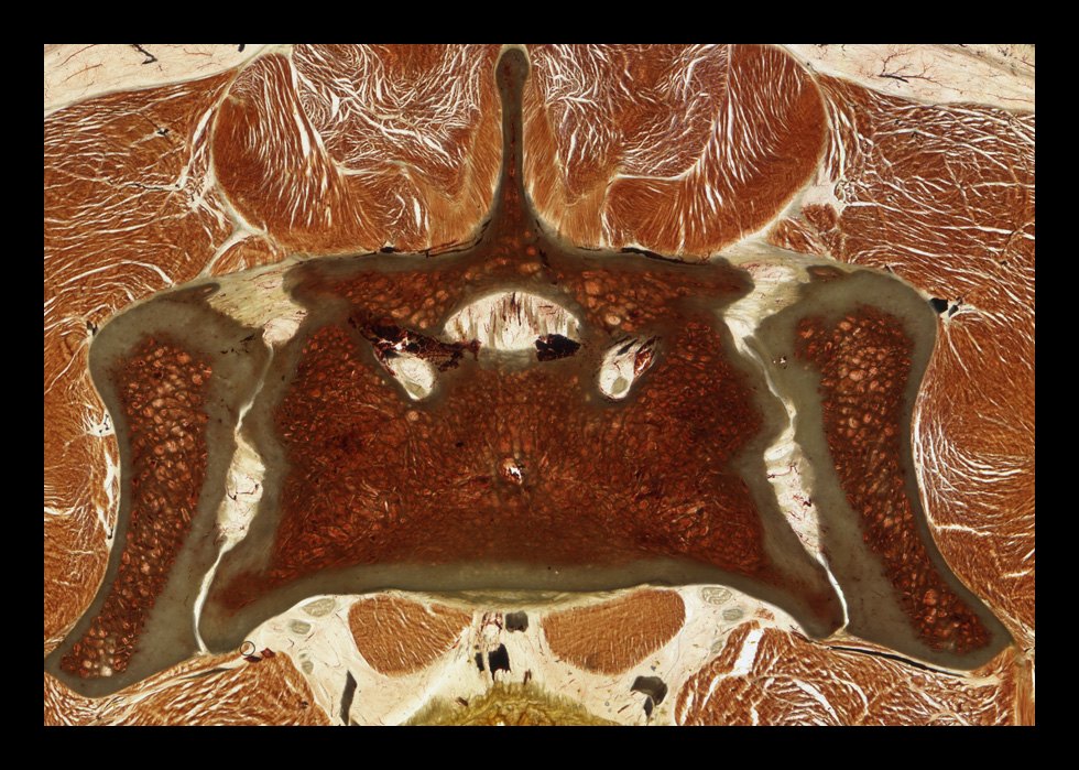
E12 epoxy-plastinated section of the canine sacroiliac region.
Figure 1.2. E12 epoxy-plastinated section of the dog sacroiliac region.

Polyester Plastination: A Powerful Tool for Neuroanatomy

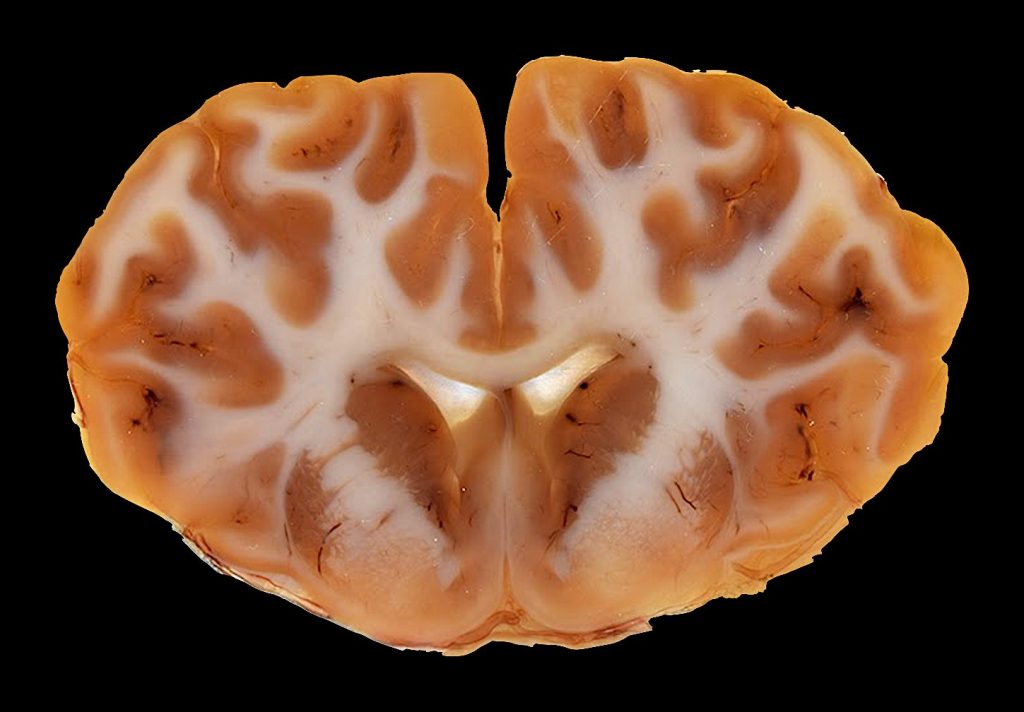
Polyester plastination is also primarily a sheet plastination technique, but it has traditionally been particularly associated with the study of the central nervous system. Polyester methods such as P35 and P40 produce firm, thin, semitransparent or transparent anatomical slices with excellent preservation of spatial relationships [1,3]. One of the major advantages of polyester in brain sections is its ability to provide a clear distinction between grey and white matter, facilitating the identification of internal neuroanatomical structures. This makes polyester-plastinated sections especially valuable for teaching neuroanatomy and for comparing anatomical sections with CT and MRI images.

Although polyester techniques can also be applied to other anatomical regions, their ability to preserve and display the internal organisation of the brain has made them particularly relevant in neurological and neuroanatomical education and research [3].
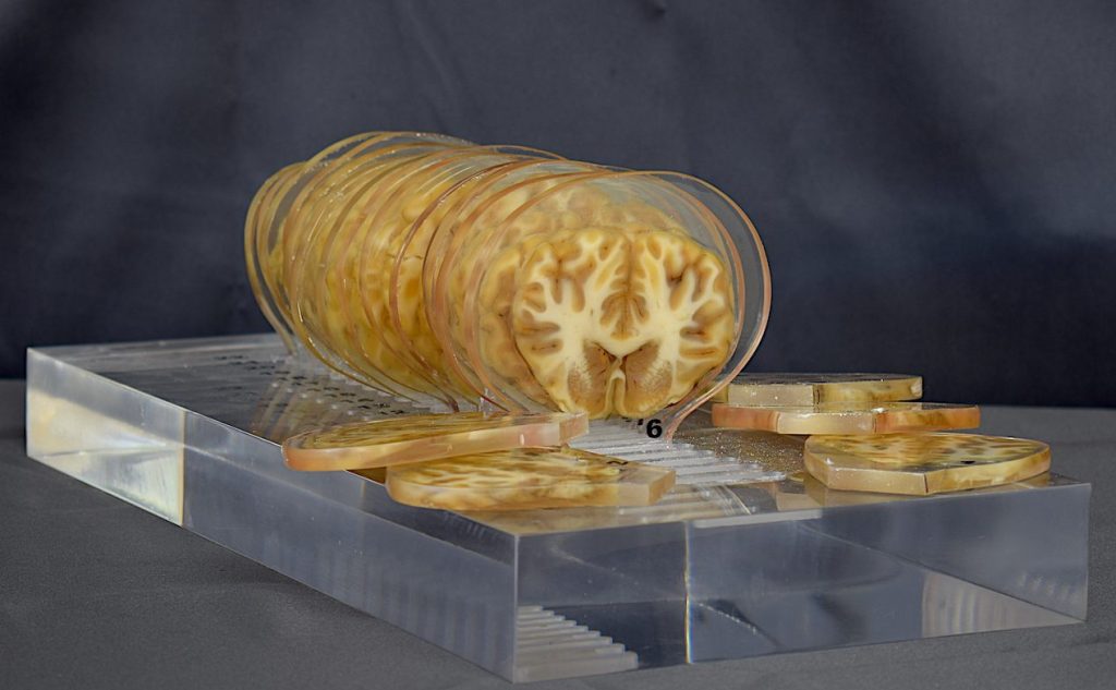
Bovine brain sections preserved by P40 polyester plastination.
Figure 1.3. Bovine brain sections preserved by P40 polyester plastination.

Choosing the Right Plastination Technique

There is therefore no single “best” plastination technique. The appropriate method depends primarily on the anatomical question and the intended use of the specimen.

Silicone plastination is generally the technique of choice when the objective is to preserve a dissected specimen in three dimensions and allow repeated handling. Epoxy plastination is particularly suited to thin transparent sections when detailed topographic anatomy and correlation with diagnostic imaging are required. Polyester plastination, although also applicable to sheet preparations, is especially valuable for neuroanatomical sections because of its ability to differentiate structures within the central nervous system.

These techniques should therefore be regarded as complementary rather than competing approaches. Together, they allow plastination to preserve anatomy at very different scales and in very different formats: from an entire organ or anatomical region that students can hold and rotate in their hands to a transparent section only a few millimetres thick that reveals the internal organisation of tissues.

The versatility of plastination lies precisely in this capacity to adapt the preservation method to the anatomical objective. By selecting the appropriate polymer and protocol, anatomical material can be transformed into durable educational and research resources while retaining the structural relationships that make real specimens so valuable for understanding anatomy.

References

  1. Sora MC, Latorre R, Baptista CAC, López-Albors O. Plastination—A scientific method for teaching and research. Anatomia, Histologia, Embryologia. 2019;48(6):526–531. https://doi.org/10.1111/ahe.12493
  2. von Hagens G, Tiedemann K, Kriz W. The current potential of plastination. Anatomy and Embryology. 1987;175(4):411–421. https://doi.org/10.1007/BF00309677
  3. Riederer BM. Plastination and its importance in teaching anatomy: Critical points for long-term preservation of human tissue. Journal of Anatomy. 2014;224(3):309–315. https://doi.org/10.1111/joa.12056
  4. Latorre R, de Jong K, Sora MC, López-Albors O, Baptista C. E12 technique: Conventional epoxy resin sheet plastination. Anatomia, Histologia, Embryologia. 2019;48(6):557–563. https://doi.org/10.1111/ahe.12507
  5. Ottone NE, Baptista CAC, Latorre R, et al. E12 sheet plastination: Techniques and applications. Clinical Anatomy. 2018;31(5):742–756. https://doi.org/10.1002/ca.23008
Sheet Plastination as a Research Tool

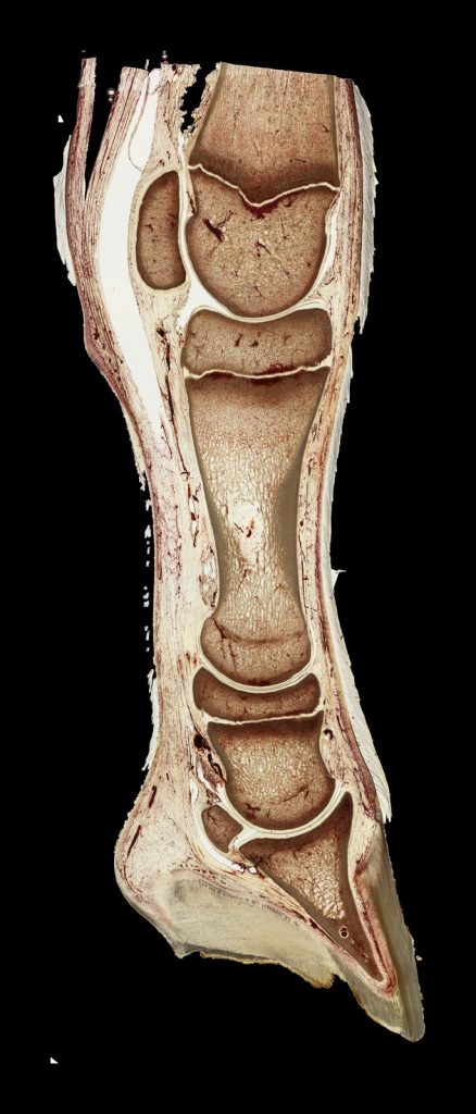
Plastination is best known as a method for preserving anatomical specimens, but it also provides a valuable material for morphological research. Thanks to the unique properties of sheet plastination, which maintains all anatomical structures in their original position, research on topographical anatomy offers multiple options for researchers and clinicians. Whereas dissection normally changes the position of structures, plastinated sections preserve the relationships between bones, muscles, connective tissues like fascia, vessels and nerves in their original position, allowing them to be examined, measured and digitised.

Epoxy-plastinated section of a foal digit, preserving relationships between bone and soft tissues.
Figure 2.1. Epoxy-plastinated section of a foal digit, preserving relationships between bone and soft tissues.

Correlating Anatomy with Medical Imaging

One important application of sheet plastination is the validation and interpretation of diagnostic images. A specimen can be imaged before sectioning, and the resulting plastinated slices can then be compared with the corresponding computed tomography (CT) or magnetic resonance imaging (MRI) planes. A valid example is the work of Steinke [1]. This author prepared a series of 800-µm-thick plastinated human body sections specifically for comparison with MRI. The study showed that these thin plastinated slices could confirm anatomical structures seen in the images and reveal important additional details. However, reliable correlation depended on accurately matching the orientation and level of the physical section with the original imaging plane. Plastination therefore provides a permanent anatomical reference for investigating the structural basis of radiological appearances.

P40-plastinated bovine brain section as an anatomical reference for imaging correlation.
Figure 2.2. P40-plastinated bovine brain section as an anatomical reference for imaging correlation.

From Serial Sections to Three-Dimensional Models

Serial plastinated sections can also be digitised, aligned and segmented to reconstruct anatomical regions in three dimensions. A good example of this is the work of Sora and co-workers [2]. A series of 1-mm thickness epoxy-plastinated sections of a human ankle were used to model bones, ligaments, fascia, the tibial nerve and the posterior tibial artery. The reconstructed anatomy was consistent with cadaveric observations. Moreover, after morphometric measurements the results did not differ significantly from those obtained from a comparison non-plastinated sample.

Additionally, such reconstructions allowed selected structures to be displayed separately or together, rotated and measured. The value of this reconstruction extends beyond visualisation: it provides a basis for analysing complex spatial relationships that may be difficult to follow through dissection or individual two-dimensional images. This single study is just an example of many other similar studies on other anatomical specimens.

Linking Macroscopic and Microscopic Anatomy

Epoxy plastination is particularly useful at the boundary between gross anatomy (macroscopy) and histology (microscopy). This mesoscopic level was explored in a detailed study of the human optic canal [3]. Using transverse, coronal and sagittal epoxy-plastinated sections from nine cadaveric heads, examined under stereoscopic and confocal microscopy, the authors traced the fibrous architecture surrounding the optic nerve, its subarachnoid space and the ophthalmic artery. The study showed that the pia and arachnoid maters merged within the optic canal and identified a dense trabecular meshwork in its orbital portion, providing an anatomical basis for reduced free communication between the intracranial subarachnoid space and the optic nerve sheath. This work illustrates how sheet plastination can preserve complex relationships within a bony compartment while supporting examination at both macroscopic and microscopic levels.

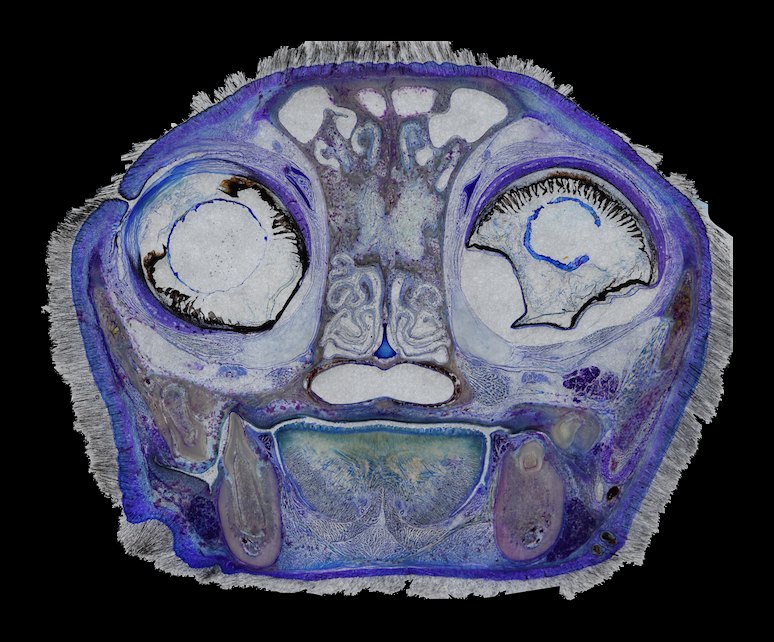
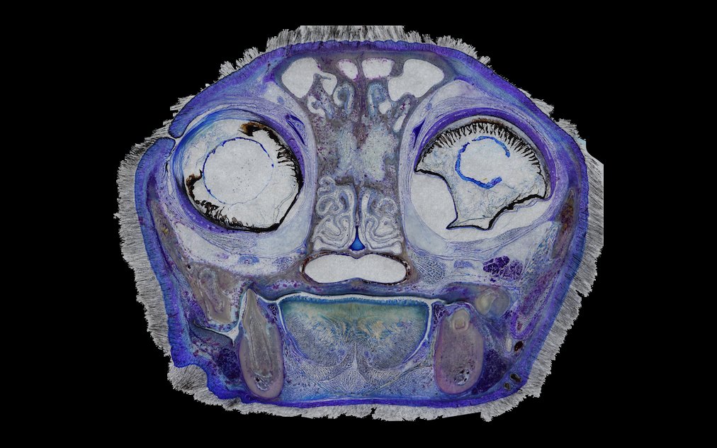
Research possibilities can be extended through staining and grinding. Staining performed before or after plastination can improve differentiation among connective tissue, cartilage, muscle and bone [4]. More recently, block-plastination techniques were used to produce sections below 300 µm with a diamond saw. Subsequent grinding was then used to reduce them to approximately 40–100 µm thickness, allowing histological staining and limited optical or fluorescence microscopy [5]. These preparations do not replace conventional histology, but they can help verify tissue identity while preserving the wider anatomical perspective.

Ultrathin epoxy-plastinated section of a cat head for detailed tissue examination.
Figure 2.3. Ultrathin epoxy-plastinated section of a cat head for detailed tissue examination.

Selective grinding also supports the Tissue Tracing Technique [5]. Instead of restricting observation to a fixed transverse, sagittal or frontal plane, researchers progressively expose a structure through a thick plastinated preparation. This makes it possible to follow curved fasciae, ligaments, tendons or joint capsules along their anatomical course.

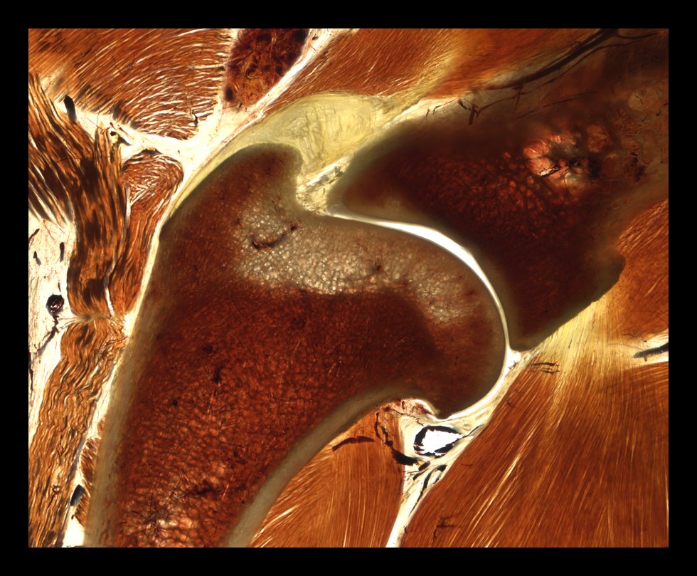
E12 epoxy-plastinated canine elbow section showing joint and periarticular structures.
Figure 2.4. E12 epoxy-plastinated canine elbow section showing joint and periarticular structures.

A Complementary Research Platform

Plastination is most informative when integrated with other methods. It can connect sectional anatomy with CT and MRI, convert serial physical sections into measurable 3D models and relate regional morphology to microscopic tissue organisation. Its limitations must also be recognised: ultrathin block plastination requires specialised cutting and grinding equipment, involves more processing steps than conventional sheet plastination, requires a previous long expertise in plastination and may take several weeks [5].

References

  1. Steinke H. Plastinated body slices for verification of magnetic resonance tomography images. Annals of Anatomy. 2001;183(3):275-281. https://doi.org/10.1016/S0940-9602(01)80234-X
  2. Sora MC, Genser-Strobl B, Radu J, Lozanoff S. Three-dimensional reconstruction of the ankle by means of ultrathin slice plastination. Clinical Anatomy. 2007;20(2):196-200. https://doi.org/10.1002/ca.20335
  3. Liugan M, Xu Z, Zhang M. Reduced free communication of the subarachnoid space within the optic canal in the human. American Journal of Ophthalmology. 2017;179:25-31. https://doi.org/10.1016/j.ajo.2017.04.012
  4. Steinke H, Rabi S, Saito T. Staining body slices before and after plastination. European Journal of Anatomy. 2008;12(1):51-55. https://eurjanat.com/v1/data/pdf/eja.08010051.pdf
  5. Sora MC, von Horst C, López-Albors O, Latorre R. Ultra-thin sectioning and grinding of epoxy plastinated tissue. Anatomia, Histologia, Embryologia. 2019;48(6):564-571. https://doi.org/10.1111/ahe.12478

How plastination has revolutionized anatomy education around the world

For centuries, the study of anatomy relied primarily on direct observation and dissection of real biological specimens. Even today, despite their unquestionable educational value, traditional specimens have important limitations: they require dedicated storage and handling facilities, may restrict students’ access to scheduled laboratory sessions, and commonly used preservation methods can raise health and safety concerns while altering the colour, texture, and overall appearance of tissues.

The development of plastination by Gunther von Hagens in the late 1970s marked a major turning point in anatomical preservation. Its significance lay not simply in extending the lifespan of anatomical specimens, but in making authentic biological material easier to handle and allowing it to move beyond the traditional dissection room into classrooms, museums, training centres, and universities worldwide [1,2].

Plastination is based on the replacement of water and part of the lipids within biological tissues by a curable polymer. The resulting specimens are dry, odourless, durable, and suitable for repeated handling in a wide variety of teaching and exhibition settings. This is central to the educational value of the technique. Whereas artificial models, whether made from resin, synthetic materials, or produced by 3D printing, typically provide standardised representations of anatomy, plastinated specimens originate from real biological material and therefore retain much of its anatomical detail and natural individual variation.

Bringing Real Anatomy Beyond the Dissection Room

The distinctive properties of plastinated specimens have had a considerable impact on the organisation of anatomy teaching. Specimens no longer need to remain immersed in preservative fluids or be restricted exclusively to the dissection laboratory. Instead, they can be incorporated into conventional classrooms, seminars, practical stations, examinations, and small-group teaching sessions. A single specimen can consequently be studied by successive cohorts of students, photographed and annotated, compared with an anatomical atlas, or examined alongside diagnostic images. This flexibility is particularly valuable in programmes with limited dissection time or restricted access to cadaveric material. Moreover, specimens requiring many hours of specialised dissection can remain available as teaching resources for years, reducing the need to reproduce complex preparations for every new course [3].

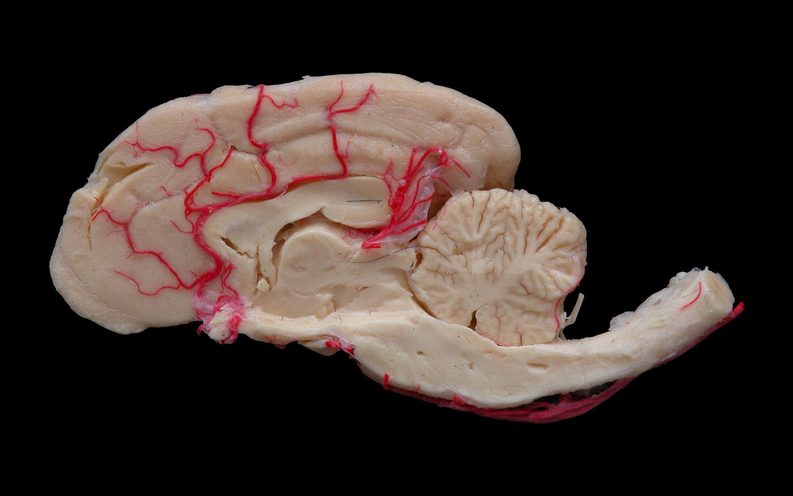
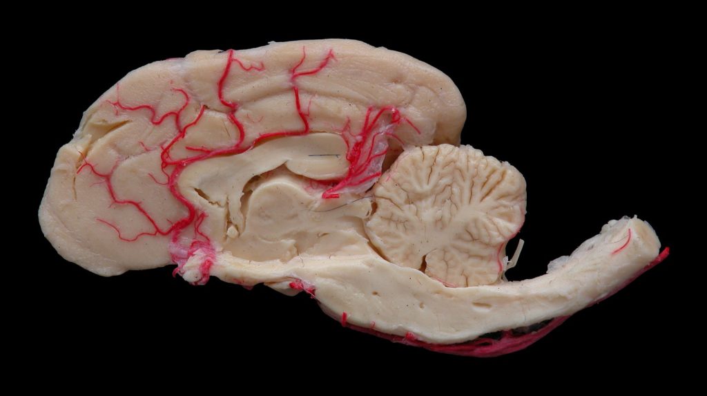
Medial view of a canine half-brain preserved by S10 silicone plastination.
Figure 3.1. Medial view of a dog half-brain preserved by S10 silicone plastination.

Understanding Anatomy in Three Dimensions

One of the most important contributions of plastination to anatomy education concerns the understanding of topographic anatomy. Genuine comprehension of an anatomical region requires more than identifying isolated structures. Students need to follow anatomical pathways, assess depth, understand relationships between adjacent structures, and mentally reconstruct three-dimensional arrangements that may be difficult to appreciate in textbook illustrations.

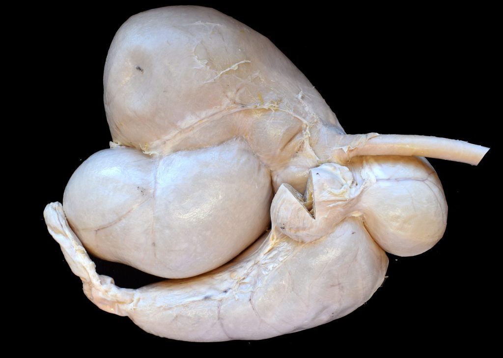
Plastinated sheep stomach for studying the three-dimensional arrangement of its compartments.
Figure 3.2. Plastinated sheep stomach for studying the three-dimensional arrangement of its compartments.

Silicone plastinated specimens can be handled, rotated, and examined from multiple perspectives, while epoxy or polyester sheet plastination produces thin anatomical sections that can be directly correlated with computed tomography (CT) and magnetic resonance imaging (MRI). In this context, the physical specimen acts as a bridge between gross anatomy and diagnostic imaging, helping students identify the anatomical structures in radiological images and follow how their relationships change across successive sections [4].

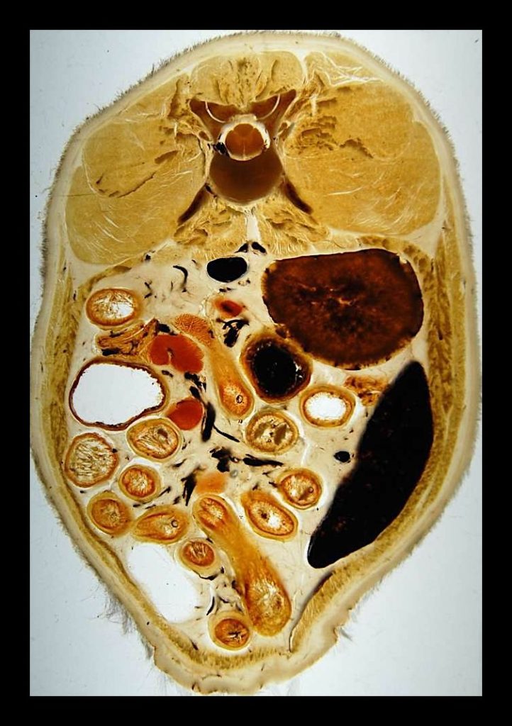
Plastinated body section for studying topographic anatomy and its relationship with diagnostic images.
Figure 3.3. Plastinated body section of dog abdomen for studying topographic anatomy and its relationship with diagnostic images.

This relationship between plastinated anatomy and medical imaging has proved particularly valuable in veterinary education. Plastinated sections, for example, have been used to correlate the anatomy of the equine tarsus with magnetic resonance images and to establish reference information on the position and dimensions of its constituent structures [5]. Other applications have extended plastination to other clinical settings. Flexible plastinated canine gastrointestinal tracts have been used to support the teaching of endoscopic anatomy hence helping the students understand the trajectory and orientation of the endoscope within the gastrointestinal tract [6].

From Universities to Schools

The international development of plastination has extended well beyond its original applications. Although initially concentrated in medical and veterinary schools, plastinated specimens are now used in a variety of healthcare programmes, postgraduate and professional training, scientific exhibitions, and museums. More recently, their educational potential has also been explored in secondary education [7].

It is worth mentioning that Discover-IN has participated in the Erasmus+ KA220-SCH OrganKits project (www.organkitsproject.eu), in which plastinated organs were incorporated into educational activities designed specifically for secondary-school students within a STEAM framework. These resources allowed students to interact directly with real anatomical material while engaging in interdisciplinary learning activities related to health and other areas of the school curriculum. Rather than using anatomical specimens only for the identification of structures in biological practices, the educational approach encouraged students to investigate questions, analyse evidence, discuss health-related problems, and draw conclusions through project-based and inquiry-oriented activities in other subjects such as mathematics, engineering, arts, physical education, etc. From our point of view, this project adds a broader educational potential to plastination: from resources traditionally associated with university anatomy laboratories to educational tools that can support scientific literacy and interdisciplinary learning in pre-university settings.

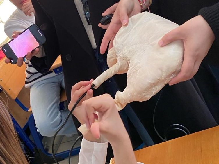
NutriHealth classroom activity using a plastinated stomach to explore digestive anatomy.
Figure 3.4. NutriHealth classroom activity using a plastinated stomach to explore digestive anatomy.

Does Plastination Improve Learning?

Describing plastination as a revolution in anatomy education does not imply that it should replace dissection. Indeed, the available evidence does not support such a conclusion. A systematic review and meta-analysis published in 2024 found that learning outcomes obtained using plastinated specimens were broadly comparable with those achieved using other anatomy teaching approaches, rather than demonstrating a consistent overall superiority of plastination. Students nevertheless showed favourable perceptions of plastinated specimens, particularly regarding their ease of use, motivational value, and usefulness for understanding anatomical structures and spatial relationships. The authors also highlighted the relatively limited number and methodological heterogeneity of comparative studies available [8]. The strongest educational argument for plastination is therefore not that it represents a universally superior teaching method, but that it provides a highly versatile form of authentic anatomical material. Its value is likely to be greatest when incorporated into a multimodal learning environment alongside dissection, diagnostic imaging, digital and 3D resources, clinical cases, and other active-learning approaches.

Extending the Life and Reach of Real Anatomy

Perhaps the most significant achievement of plastination has been to make authentic anatomy more accessible, durable, and adaptable to different educational environments. A specimen that might once have been available only for a limited period can become a long-term educational resource. A complex dissection requiring many hours of preparation can be studied by successive generations of students. An anatomical section can be placed directly alongside an MRI or CT image, allowing learners to understand precisely which structures underlie the clinical image. And, anatomical material once largely confined to specialised university laboratories can now be incorporated into teaching environments far beyond the traditional dissection room.

Thus, plastination has not eliminated, and should not be expected to eliminate, the need to explore anatomy through other methods. What it has fundamentally changed is where, when, and how authentic anatomical specimens can be studied. In that sense, the real revolution of plastination is not simply that anatomical specimens can be preserved for longer. It is that real anatomy can travel further, be handled more easily, and become accessible to many more learners. This capacity to extend the anatomical specimen beyond the boundaries of the traditional laboratory is perhaps plastination’s most enduring contribution to anatomy education worldwide.

References

  1. von Hagens G, Tiedemann K, Kriz W. The current potential of plastination. Anatomy and Embryology. 1987;175(4):411–421. https://doi.org/10.1007/BF00309677
  2. Sora MC, Latorre R, Baptista CAC, López-Albors O. Plastination—A scientific method for teaching and research. Anatomia, Histologia, Embryologia. 2019;48(6):526–531. https://doi.org/10.1111/ahe.12493
  3. Riederer BM. Plastination and its importance in teaching anatomy: Critical points for long-term preservation of human tissue. Journal of Anatomy. 2014;224(3):309–315. https://doi.org/10.1111/joa.12056
  4. Ottone NE, Baptista CAC, Latorre R, et al. E12 sheet plastination: Techniques and applications. Clinical Anatomy. 2018;31(5):742–756. https://doi.org/10.1002/ca.23008
  5. Latorre R, Arencibia A, Gil F, et al. Correlation of magnetic resonance images with anatomic features of the equine tarsus. American Journal of Veterinary Research. 2006;67(5):756–761. https://doi.org/10.2460/ajvr.67.5.756
  6. Janick L, DeNovo RC, Henry RW. Plastinated canine gastrointestinal tracts used to facilitate teaching of endoscopic technique and anatomy. Acta Anatomica. 1997;158(1):48–53. https://doi.org/10.1159/000147910
  7. Delgado-Ruiz MC, Bernal-Sánchez R, López-Albors O, Latorre-Reviriego R. Órganos plastinados: recursos para una propuesta de innovación educativa STEM en Educación Secundaria. RiiTE. 2023;15:103–119. https://doi.org/10.6018/riite.575831
  8. Goh JSK, Chandrasekaran R, Sirasanagandla SR, Acharyya S, Mogali SR. Efficacy of plastinated specimens in anatomy education: A systematic review and meta-analysis. Anatomical Sciences Education. 2024;17(4):712–721. https://doi.org/10.1002/ase.2424

OrganKits for Secondary Schools

www.organkitsproject.eu

OrganKits is a European Erasmus+ project coordinated by the University of Murcia that has developed, implemented and evaluated an educational model for STEAM teaching in secondary education. Its approach integrates plastinated organs with interdisciplinary activities that connect anatomy and health with social and environmental issues.

The project addresses a specific need: providing schools with real, durable anatomical materials that can be handled repeatedly, together with a methodology for using them across different subjects. On this basis, the OrganKits Educational Model combines anatomical specimens, project-based learning activities and teaching support resources.

Six Educational Kits Connecting Health and Society

The model is organised into six thematic areas, each known as an OrganKit:

  • CardioHealth & Emotions: cardiovascular health and emotions.
  • NeumoHealth & Environment: respiratory health and the environment.
  • NutriHealth & Wellness: nutrition and well-being.
  • MentalHealth & Mindfulness: mental health and mindfulness.
  • SportsHealth & Dependence: health, sport and dependence.
  • ReproHealth & Gender: reproductive health and gender.

Each OrganKit brings together anatomical materials, a STEAM learning framework and resources for classroom implementation. Plastinated organs form the physical core of the activities and serve as a key source of motivation. Educational guides and teacher guides structure the learning process, while atlases, digital resources and interdisciplinary implementation maps support classroom delivery.

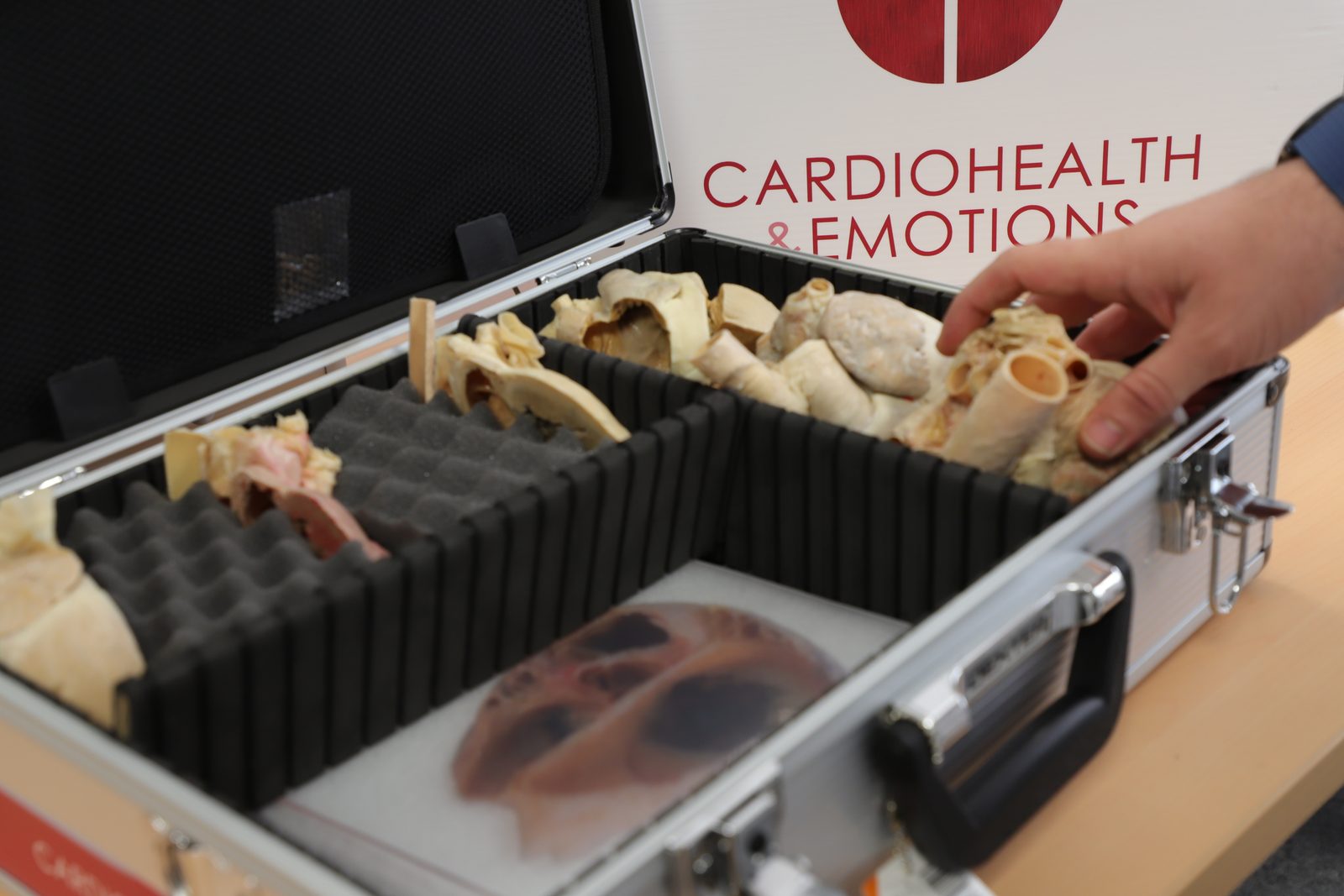
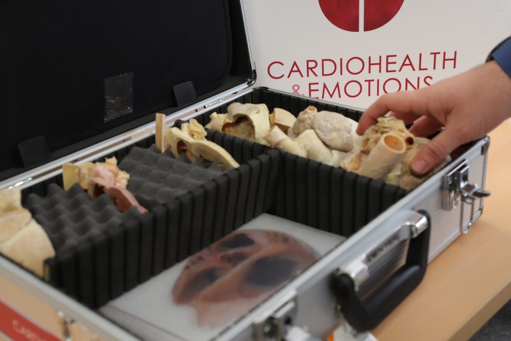
CardioHealth carrying case with plastinated specimens and supporting teaching materials.
Figure 4.1. CardioHealth OrganKit, briefcase with plastinated specimens.

The plastinated specimens, obtained from pigs, cattle and sheep, allow students to study real anatomical structures and use them as a starting point to investigate issues relevant to everyday life. The pedagogical innovation lies in extending their use across STEAM subjects, connecting anatomical content with activities and questions that draw on several disciplines.

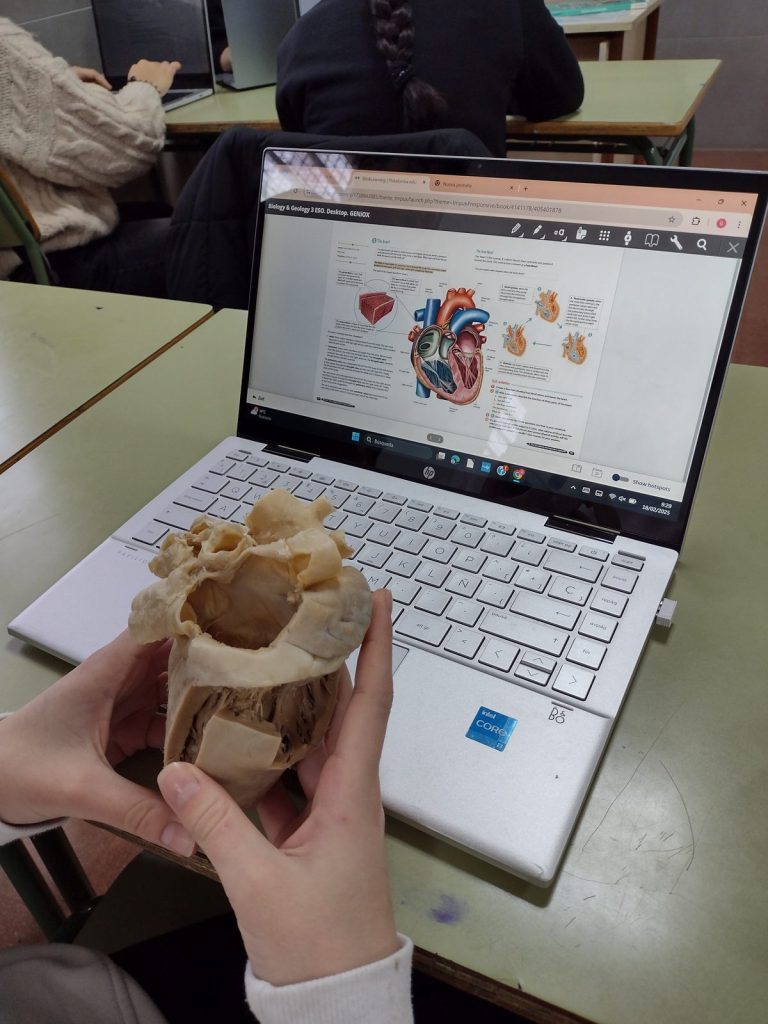
CardioHealth activity combining a real anatomical specimen with digital resources.
Figure 4.2. CardioHealth activity combining a real anatomical specimen with digital resources.

Materials and Resources Developed

The project produced 479 plastinated organs, 95 more than initially planned. Six complete educational packages were developed, available in Spanish, English, Italian, Greek and Turkish. Together, these materials comprise 30 educational guides, 30 teacher guides, more than 100 digital educational resources and six STEAM learning pathway maps. They also include six anatomical atlases accessible through QR codes.

These materials allow different levels of adoption. A school can select an individual activity, work with one OrganKit or implement a broader programme, adapting the resources to its curriculum, the subjects involved and students’ needs.

NeumoHealth practical activity exploring respiratory anatomy with plastinated organs.
Figure 4.3. NeumoHealth practical activity exploring respiratory anatomy with plastinated organs.

Findings from the Educational Evaluation

Students particularly valued the opportunity to observe and handle real organs, reporting that the experience supported their understanding and made the activities more engaging. Teachers highlighted the quality and ease of handling of the specimens, the usefulness of the guides and their ability to capture students’ attention. Focus groups reported perceived improvements in motivation, participation and three-dimensional understanding of anatomy. These findings support the educational usefulness and acceptance of the model in the participating settings.

Inclusion and Transfer to Other Settings

The project also explored applications in inclusive education. Experiences were documented with students with visual impairments and those with attention deficit and/or hyperactivity disorders. The resources were used at the Çağla Pınar Rehabilitation Centre in Türkiye and in workshops for young people experiencing social vulnerability at the Museum of Science and Water in Murcia. Following the project’s completion, they were also used in the hospital classrooms of the Virgen de la Arrixaca University Clinical Hospital.

Outreach activities extended to three science museums. The permanent exhibition at the Museum of Science and Water in Murcia received more than 10,000 visitors during its first three months. The project website also recorded 10,750 unique visitors, and the International Conference on OrganKits brought together 77 participants. The project’s final report received an evaluation score of 93 out of 100.

Discover-IN’s Contribution

Discover-IN participated as the technical partner responsible for developing and producing the plastinated specimens. Its expertise in anatomical preservation provided the materials on which the educational partners built and implemented their teaching activities.

This contribution was essential to meeting the project’s practical requirements: preserving anatomical detail, enabling repeated handling and facilitating storage and transport between schools. The production of durable specimens, organised into carrying cases, supported their use throughout the implementation cycles and their subsequent loan to associated schools.

The production process also incorporated sustainability measures, including reduced formaldehyde use, acetone recovery and recycling, the selection of local suppliers and the use of recyclable aluminium carrying cases.

Discover-IN’s contribution lies in applying its specialist knowledge of plastination to the needs of secondary education. In collaboration with the University of Murcia and the educational partners, this technical expertise enabled anatomical specimens to become the foundation of an educational model combining reusable materials, structured activities and opportunities for adaptation to other learning settings.