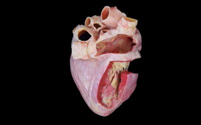
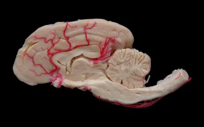
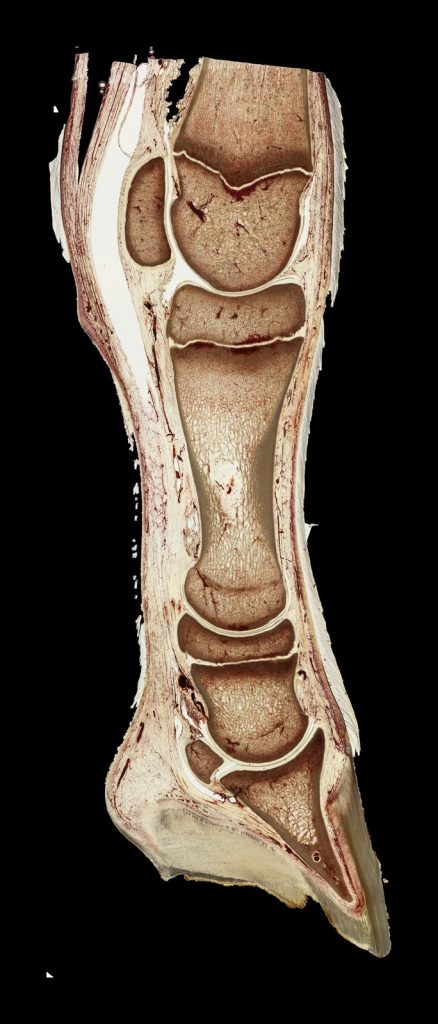
Plastination is best known as a method for preserving anatomical specimens, but it also provides a valuable material for morphological research. Thanks to the unique properties of sheet plastination, which maintains all anatomical structures in their original position, research on topographical anatomy offers multiple options for researchers and clinicians. Whereas dissection normally changes the position of structures, plastinated sections preserve the relationships between bones, muscles, connective tissues like fascia, vessels and nerves in their original position, allowing them to be examined, measured and digitised.
Correlating Anatomy with Medical Imaging
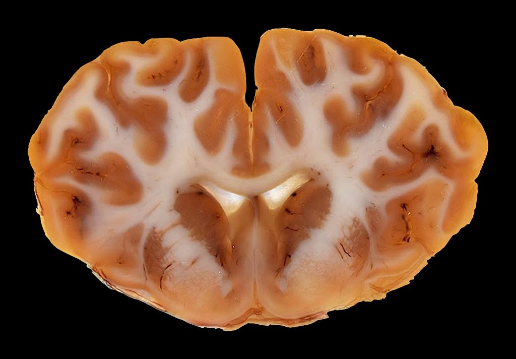
One important application of sheet plastination is the validation and interpretation of diagnostic images. A specimen can be imaged before sectioning, and the resulting plastinated slices can then be compared with the corresponding computed tomography (CT) or magnetic resonance imaging (MRI) planes. A valid example is the work of Steinke [1]. This author prepared a series of 800-µm-thick plastinated human body sections specifically for comparison with MRI. The study showed that these thin plastinated slices could confirm anatomical structures seen in the images and reveal important additional details. However, reliable correlation depended on accurately matching the orientation and level of the physical section with the original imaging plane. Plastination therefore provides a permanent anatomical reference for investigating the structural basis of radiological appearances.
From Serial Sections to Three-Dimensional Models
Serial plastinated sections can also be digitised, aligned and segmented to reconstruct anatomical regions in three dimensions. A good example of this is the work of Sora and co-workers [2]. A series of 1-mm thickness epoxy-plastinated sections of a human ankle were used to model bones, ligaments, fascia, the tibial nerve and the posterior tibial artery. The reconstructed anatomy was consistent with cadaveric observations. Moreover, after morphometric measurements the results did not differ significantly from those obtained from a comparison non-plastinated sample.
Additionally, such reconstructions allowed selected structures to be displayed separately or together, rotated and measured. The value of this reconstruction extends beyond visualisation: it provides a basis for analysing complex spatial relationships that may be difficult to follow through dissection or individual two-dimensional images. This single study is just an example of many other similar studies on other anatomical specimens.
Linking Macroscopic and Microscopic Anatomy
Epoxy plastination is particularly useful at the boundary between gross anatomy (macroscopy) and histology (microscopy). This mesoscopic level was explored in a detailed study of the human optic canal [3]. Using transverse, coronal and sagittal epoxy-plastinated sections from nine cadaveric heads, examined under stereoscopic and confocal microscopy, the authors traced the fibrous architecture surrounding the optic nerve, its subarachnoid space and the ophthalmic artery. The study showed that the pia and arachnoid maters merged within the optic canal and identified a dense trabecular meshwork in its orbital portion, providing an anatomical basis for reduced free communication between the intracranial subarachnoid space and the optic nerve sheath. This work illustrates how sheet plastination can preserve complex relationships within a bony compartment while supporting examination at both macroscopic and microscopic levels.
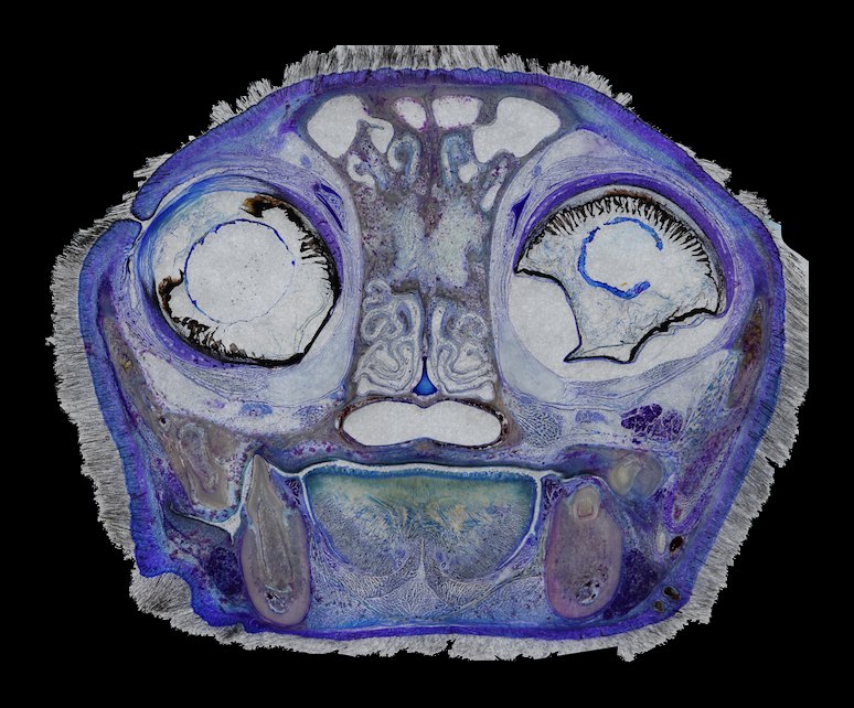
Research possibilities can be extended through staining and grinding. Staining performed before or after plastination can improve differentiation among connective tissue, cartilage, muscle and bone [4]. More recently, block-plastination techniques were used to produce sections below 300 µm with a diamond saw. Subsequent grinding was then used to reduce them to approximately 40–100 µm thickness, allowing histological staining and limited optical or fluorescence microscopy [5]. These preparations do not replace conventional histology, but they can help verify tissue identity while preserving the wider anatomical perspective.
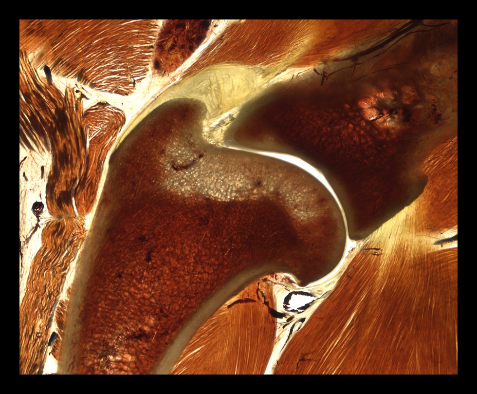
Selective grinding also supports the Tissue Tracing Technique [5]. Instead of restricting observation to a fixed transverse, sagittal or frontal plane, researchers progressively expose a structure through a thick plastinated preparation. This makes it possible to follow curved fasciae, ligaments, tendons or joint capsules along their anatomical course.
A Complementary Research Platform
Plastination is most informative when integrated with other methods. It can connect sectional anatomy with CT and MRI, convert serial physical sections into measurable 3D models and relate regional morphology to microscopic tissue organisation. Its limitations must also be recognised: ultrathin block plastination requires specialised cutting and grinding equipment, involves more processing steps than conventional sheet plastination, requires a previous long expertise in plastination and may take several weeks [5].
References
- Steinke H. Plastinated body slices for verification of magnetic resonance tomography images. Annals of Anatomy. 2001;183(3):275-281. https://doi.org/10.1016/S0940-9602(01)80234-X
- Sora MC, Genser-Strobl B, Radu J, Lozanoff S. Three-dimensional reconstruction of the ankle by means of ultrathin slice plastination. Clinical Anatomy. 2007;20(2):196-200. https://doi.org/10.1002/ca.20335
- Liugan M, Xu Z, Zhang M. Reduced free communication of the subarachnoid space within the optic canal in the human. American Journal of Ophthalmology. 2017;179:25-31. https://doi.org/10.1016/j.ajo.2017.04.012
- Steinke H, Rabi S, Saito T. Staining body slices before and after plastination. European Journal of Anatomy. 2008;12(1):51-55. https://eurjanat.com/v1/data/pdf/eja.08010051.pdf
- Sora MC, von Horst C, López-Albors O, Latorre R. Ultra-thin sectioning and grinding of epoxy plastinated tissue. Anatomia, Histologia, Embryologia. 2019;48(6):564-571. https://doi.org/10.1111/ahe.12478