Plastination is not a single preservation procedure, but rather a family of techniques based on a common principle: the replacement of water and part of the lipids within biological tissues by a curable polymer. Once this polymer has been introduced into the tissues and subsequently hardened, the resulting anatomical specimens are dry, odourless, durable, and suitable for long-term handling, teaching, research, or exhibition [1,2].
Although different plastination protocols and polymers have been developed since the technique was introduced by Gunther von Hagens in the late 1970s, three major approaches have become particularly important: silicone, epoxy, and polyester plastination. Each produces specimens with different physical and optical properties and, consequently, each is particularly suited to specific anatomical applications.
The Common Principles of Plastination
Regardless of the polymer used, conventional plastination follows a series of fundamental steps. First, the specimen is prepared and, in most cases, fixed to preserve its anatomical structures. The tissue is then dehydrated, usually with acetone, removing the water contained within it and part of its lipids. The central step of the process is forced impregnation. Under vacuum, the acetone within the specimen progressively vaporises and is replaced by a liquid polymer. The impregnated specimen is finally cured or hardened, transforming the polymer into a stable material permanently incorporated within the tissues [1]. The choice of polymer is crucial because it determines many of the final characteristics of the plastinated specimen, including its flexibility, transparency, mechanical resistance, and suitability for either three-dimensional preparations or anatomical slices.
Silicone Plastination: Preserving Anatomy in Three Dimensions
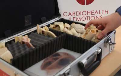
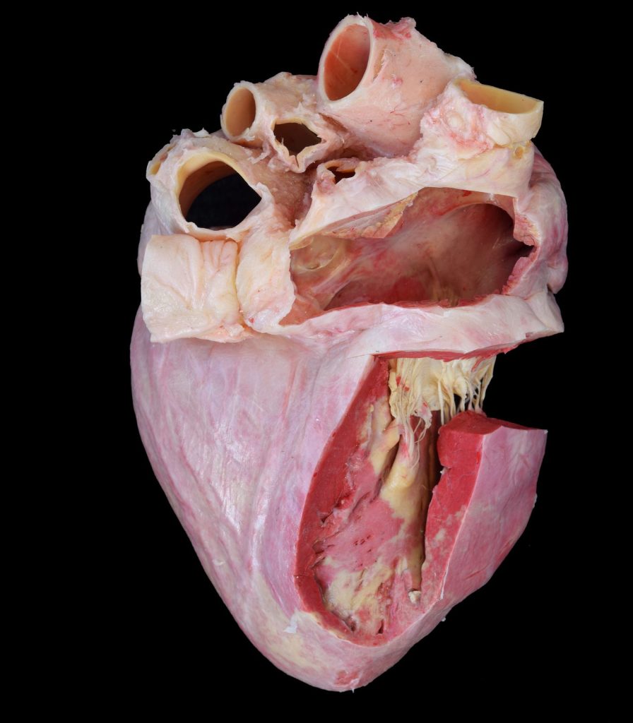
Silicone plastination is the most widely used plastination technique and is particularly suitable for preserving whole organs, body parts, dissected anatomical regions, and other three-dimensional specimens [1–3]. After forced impregnation with silicone, the specimen is positioned appropriately and the polymer is cured. The resulting preparations are generally opaque or only slightly translucent and retain a relatively natural appearance. Depending on the protocol and the tissues involved, they may also preserve a certain degree of flexibility.
One of the main advantages of silicone plastination is that it maintains the three-dimensional relationships between anatomical structures while producing specimens that can be handled repeatedly without the need for immersion in preservative fluids. This makes silicone plastinates especially useful for gross anatomy teaching, where students need to identify structures, follow vessels and nerves, understand anatomical relationships, and examine a specimen from different perspectives. For this reason, silicone plastination has become particularly valuable for the long-term preservation of carefully dissected specimens. A complex anatomical preparation that may require many hours of dissection can remain available for successive generations of students and can be used in practical classes, examinations, demonstrations, and exhibitions [2,3].
Epoxy Plastination: Anatomy in Transparent Sections
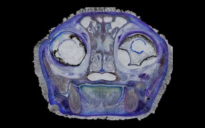
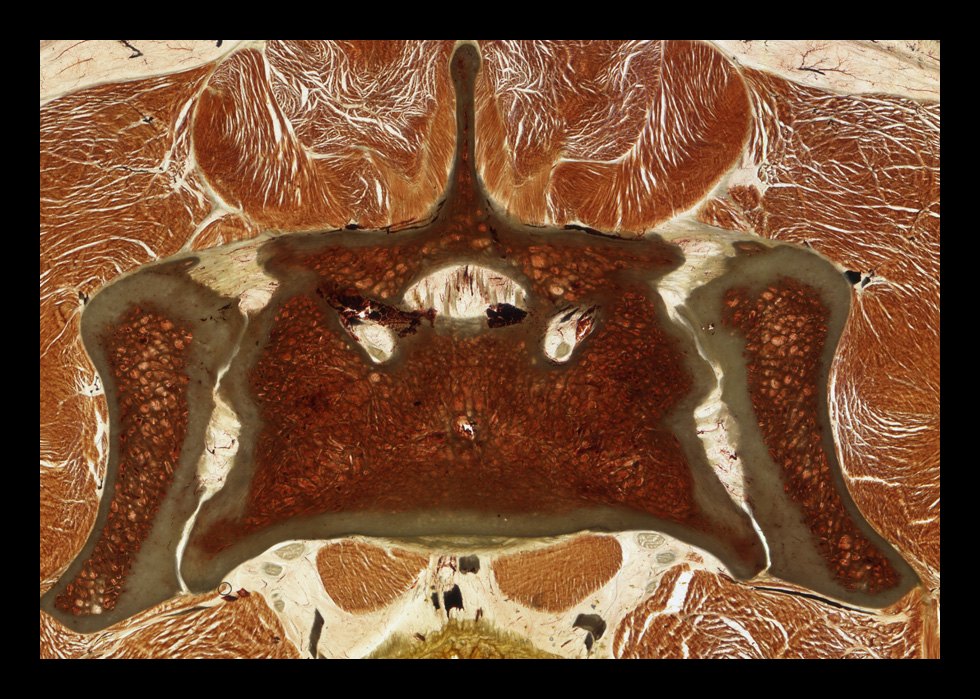
Epoxy plastination has a very different purpose. Rather than preserving complete three-dimensional preparations, epoxy resins are mainly used to produce thin, transparent anatomical slices. In epoxy sheet plastination, frozen anatomical material is sectioned into thin slices, commonly only a few millimetres thick. Following dehydration and forced impregnation with epoxy resin, the sections are cast and cured to produce firm, transparent preparations with a high degree of anatomical detail [1,4]. Their transparency is one of their most important characteristics. Structures can be examined within their original topographical context without destroying the relationships between tissues. Depending on section thickness details ranging from gross anatomical structures to considerably thinner morphological features can be investigated [4].
Epoxy plastination is therefore especially valuable for sectional and topographic anatomy and for correlating physical anatomical sections with diagnostic imaging techniques such as computed tomography (CT) and magnetic resonance imaging (MRI). The technique has consequently become an important tool not only for teaching but also for anatomical and morphological research [4,5].
Polyester Plastination: A Powerful Tool for Neuroanatomy
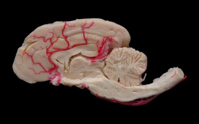
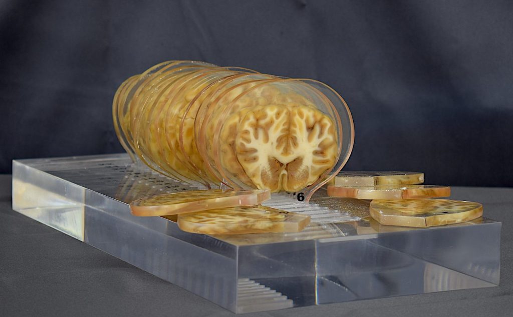
Polyester plastination is also primarily a sheet plastination technique, but it has traditionally been particularly associated with the study of the central nervous system. Polyester methods such as P35 and P40 produce firm, thin, semitransparent or transparent anatomical slices with excellent preservation of spatial relationships [1,3]. One of the major advantages of polyester in brain sections is its ability to provide a clear distinction between grey and white matter, facilitating the identification of internal neuroanatomical structures. This makes polyester-plastinated sections especially valuable for teaching neuroanatomy and for comparing anatomical sections with CT and MRI images.
Although polyester techniques can also be applied to other anatomical regions, their ability to preserve and display the internal organisation of the brain has made them particularly relevant in neurological and neuroanatomical education and research [3].
Choosing the Right Plastination Technique
There is therefore no single “best” plastination technique. The appropriate method depends primarily on the anatomical question and the intended use of the specimen.
Silicone plastination is generally the technique of choice when the objective is to preserve a dissected specimen in three dimensions and allow repeated handling. Epoxy plastination is particularly suited to thin transparent sections when detailed topographic anatomy and correlation with diagnostic imaging are required. Polyester plastination, although also applicable to sheet preparations, is especially valuable for neuroanatomical sections because of its ability to differentiate structures within the central nervous system.
These techniques should therefore be regarded as complementary rather than competing approaches. Together, they allow plastination to preserve anatomy at very different scales and in very different formats: from an entire organ or anatomical region that students can hold and rotate in their hands to a transparent section only a few millimetres thick that reveals the internal organisation of tissues.
The versatility of plastination lies precisely in this capacity to adapt the preservation method to the anatomical objective. By selecting the appropriate polymer and protocol, anatomical material can be transformed into durable educational and research resources while retaining the structural relationships that make real specimens so valuable for understanding anatomy.
References
- Sora MC, Latorre R, Baptista CAC, López-Albors O. Plastination—A scientific method for teaching and research. Anatomia, Histologia, Embryologia. 2019;48(6):526–531. https://doi.org/10.1111/ahe.12493
- von Hagens G, Tiedemann K, Kriz W. The current potential of plastination. Anatomy and Embryology. 1987;175(4):411–421. https://doi.org/10.1007/BF00309677
- Riederer BM. Plastination and its importance in teaching anatomy: Critical points for long-term preservation of human tissue. Journal of Anatomy. 2014;224(3):309–315. https://doi.org/10.1111/joa.12056
- Latorre R, de Jong K, Sora MC, López-Albors O, Baptista C. E12 technique: Conventional epoxy resin sheet plastination. Anatomia, Histologia, Embryologia. 2019;48(6):557–563. https://doi.org/10.1111/ahe.12507
- Ottone NE, Baptista CAC, Latorre R, et al. E12 sheet plastination: Techniques and applications. Clinical Anatomy. 2018;31(5):742–756. https://doi.org/10.1002/ca.23008