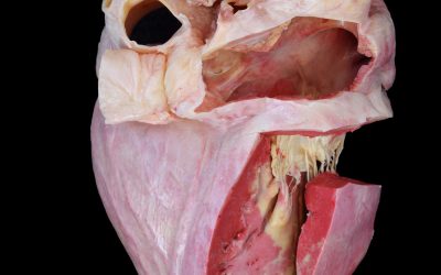
For centuries, the study of anatomy relied primarily on direct observation and dissection of real biological specimens. Even today, despite their unquestionable educational value, traditional specimens have important limitations: they require dedicated storage and handling facilities, may restrict students’ access to scheduled laboratory sessions, and commonly used preservation methods can raise health and safety concerns while altering the colour, texture, and overall appearance of tissues.
The development of plastination by Gunther von Hagens in the late 1970s marked a major turning point in anatomical preservation. Its significance lay not simply in extending the lifespan of anatomical specimens, but in making authentic biological material easier to handle and allowing it to move beyond the traditional dissection room into classrooms, museums, training centres, and universities worldwide [1,2].
Plastination is based on the replacement of water and part of the lipids within biological tissues by a curable polymer. The resulting specimens are dry, odourless, durable, and suitable for repeated handling in a wide variety of teaching and exhibition settings. This is central to the educational value of the technique. Whereas artificial models, whether made from resin, synthetic materials, or produced by 3D printing, typically provide standardised representations of anatomy, plastinated specimens originate from real biological material and therefore retain much of its anatomical detail and natural individual variation.
Bringing Real Anatomy Beyond the Dissection Room
The distinctive properties of plastinated specimens have had a considerable impact on the organisation of anatomy teaching. Specimens no longer need to remain immersed in preservative fluids or be restricted exclusively to the dissection laboratory. Instead, they can be incorporated into conventional classrooms, seminars, practical stations, examinations, and small-group teaching sessions. A single specimen can consequently be studied by successive cohorts of students, photographed and annotated, compared with an anatomical atlas, or examined alongside diagnostic images. This flexibility is particularly valuable in programmes with limited dissection time or restricted access to cadaveric material. Moreover, specimens requiring many hours of specialised dissection can remain available as teaching resources for years, reducing the need to reproduce complex preparations for every new course [3].
Understanding Anatomy in Three Dimensions
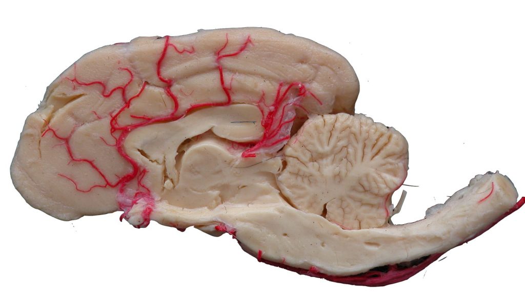
One of the most important contributions of plastination to anatomy education concerns the understanding of topographic anatomy. Genuine comprehension of an anatomical region requires more than identifying isolated structures. Students need to follow anatomical pathways, assess depth, understand relationships between adjacent structures, and mentally reconstruct three-dimensional arrangements that may be difficult to appreciate in textbook illustrations.
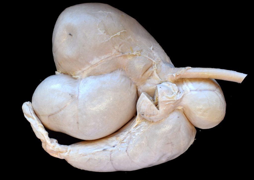
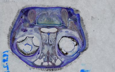
Silicone plastinated specimens can be handled, rotated, and examined from multiple perspectives, while epoxy or polyester sheet plastination produces thin anatomical sections that can be directly correlated with computed tomography (CT) and magnetic resonance imaging (MRI). In this context, the physical specimen acts as a bridge between gross anatomy and diagnostic imaging, helping students identify the anatomical structures in radiological images and follow how their relationships change across successive sections [4].
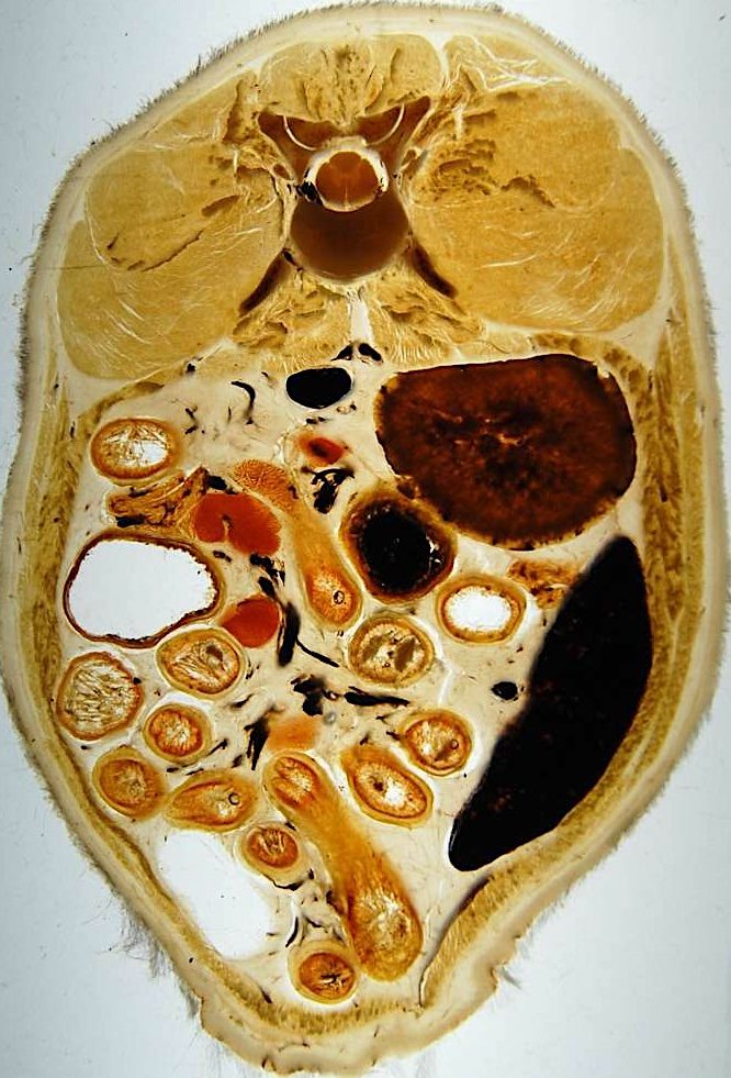
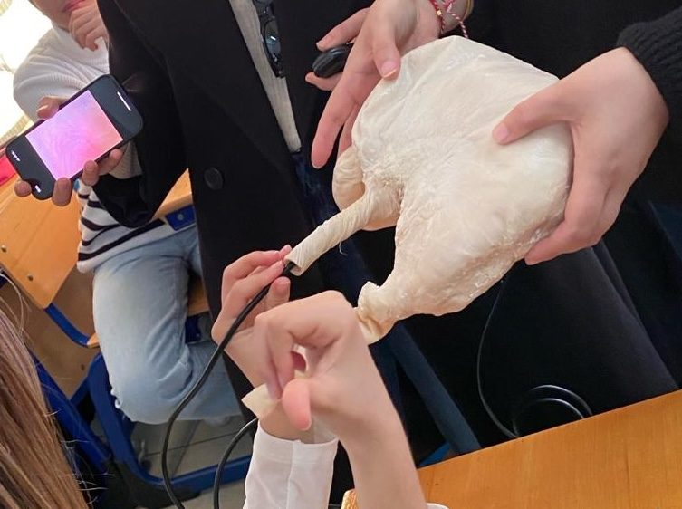
This relationship between plastinated anatomy and medical imaging has proved particularly valuable in veterinary education. Plastinated sections, for example, have been used to correlate the anatomy of the equine tarsus with magnetic resonance images and to establish reference information on the position and dimensions of its constituent structures [5]. Other applications have extended plastination to other clinical settings. Flexible plastinated canine gastrointestinal tracts have been used to support the teaching of endoscopic anatomy hence helping the students understand the trajectory and orientation of the endoscope within the gastrointestinal tract [6].
From Universities to Schools
The international development of plastination has extended well beyond its original applications. Although initially concentrated in medical and veterinary schools, plastinated specimens are now used in a variety of healthcare programmes, postgraduate and professional training, scientific exhibitions, and museums. More recently, their educational potential has also been explored in secondary education [7].
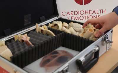
It is worth mentioning that Discover-IN has participated in the Erasmus+ KA220-SCH OrganKits project (www.organkitsproject.eu), in which plastinated organs were incorporated into educational activities designed specifically for secondary-school students within a STEAM framework. These resources allowed students to interact directly with real anatomical material while engaging in interdisciplinary learning activities related to health and other areas of the school curriculum. Rather than using anatomical specimens only for the identification of structures in biological practices, the educational approach encouraged students to investigate questions, analyse evidence, discuss health-related problems, and draw conclusions through project-based and inquiry-oriented activities in other subjects such as mathematics, engineering, arts, physical education, etc. From our point of view, this project adds a broader educational potential to plastination: from resources traditionally associated with university anatomy laboratories to educational tools that can support scientific literacy and interdisciplinary learning in pre-university settings.
Does Plastination Improve Learning?
Describing plastination as a revolution in anatomy education does not imply that it should replace dissection. Indeed, the available evidence does not support such a conclusion. A systematic review and meta-analysis published in 2024 found that learning outcomes obtained using plastinated specimens were broadly comparable with those achieved using other anatomy teaching approaches, rather than demonstrating a consistent overall superiority of plastination. Students nevertheless showed favourable perceptions of plastinated specimens, particularly regarding their ease of use, motivational value, and usefulness for understanding anatomical structures and spatial relationships. The authors also highlighted the relatively limited number and methodological heterogeneity of comparative studies available [8]. The strongest educational argument for plastination is therefore not that it represents a universally superior teaching method, but that it provides a highly versatile form of authentic anatomical material. Its value is likely to be greatest when incorporated into a multimodal learning environment alongside dissection, diagnostic imaging, digital and 3D resources, clinical cases, and other active-learning approaches.
Extending the Life and Reach of Real Anatomy
Perhaps the most significant achievement of plastination has been to make authentic anatomy more accessible, durable, and adaptable to different educational environments. A specimen that might once have been available only for a limited period can become a long-term educational resource. A complex dissection requiring many hours of preparation can be studied by successive generations of students. An anatomical section can be placed directly alongside an MRI or CT image, allowing learners to understand precisely which structures underlie the clinical image. And, anatomical material once largely confined to specialised university laboratories can now be incorporated into teaching environments far beyond the traditional dissection room.
Thus, plastination has not eliminated, and should not be expected to eliminate, the need to explore anatomy through other methods. What it has fundamentally changed is where, when, and how authentic anatomical specimens can be studied. In that sense, the real revolution of plastination is not simply that anatomical specimens can be preserved for longer. It is that real anatomy can travel further, be handled more easily, and become accessible to many more learners. This capacity to extend the anatomical specimen beyond the boundaries of the traditional laboratory is perhaps plastination’s most enduring contribution to anatomy education worldwide.
References
- von Hagens G, Tiedemann K, Kriz W. The current potential of plastination. Anatomy and Embryology. 1987;175(4):411–421. https://doi.org/10.1007/BF00309677
- Sora MC, Latorre R, Baptista CAC, López-Albors O. Plastination—A scientific method for teaching and research. Anatomia, Histologia, Embryologia. 2019;48(6):526–531. https://doi.org/10.1111/ahe.12493
- Riederer BM. Plastination and its importance in teaching anatomy: Critical points for long-term preservation of human tissue. Journal of Anatomy. 2014;224(3):309–315. https://doi.org/10.1111/joa.12056
- Ottone NE, Baptista CAC, Latorre R, et al. E12 sheet plastination: Techniques and applications. Clinical Anatomy. 2018;31(5):742–756. https://doi.org/10.1002/ca.23008
- Latorre R, Arencibia A, Gil F, et al. Correlation of magnetic resonance images with anatomic features of the equine tarsus. American Journal of Veterinary Research. 2006;67(5):756–761. https://doi.org/10.2460/ajvr.67.5.756
- Janick L, DeNovo RC, Henry RW. Plastinated canine gastrointestinal tracts used to facilitate teaching of endoscopic technique and anatomy. Acta Anatomica. 1997;158(1):48–53. https://doi.org/10.1159/000147910
- Delgado-Ruiz MC, Bernal-Sánchez R, López-Albors O, Latorre-Reviriego R. Órganos plastinados: recursos para una propuesta de innovación educativa STEM en Educación Secundaria. RiiTE. 2023;15:103–119. https://doi.org/10.6018/riite.575831
- Goh JSK, Chandrasekaran R, Sirasanagandla SR, Acharyya S, Mogali SR. Efficacy of plastinated specimens in anatomy education: A systematic review and meta-analysis. Anatomical Sciences Education. 2024;17(4):712–721. https://doi.org/10.1002/ase.2424