
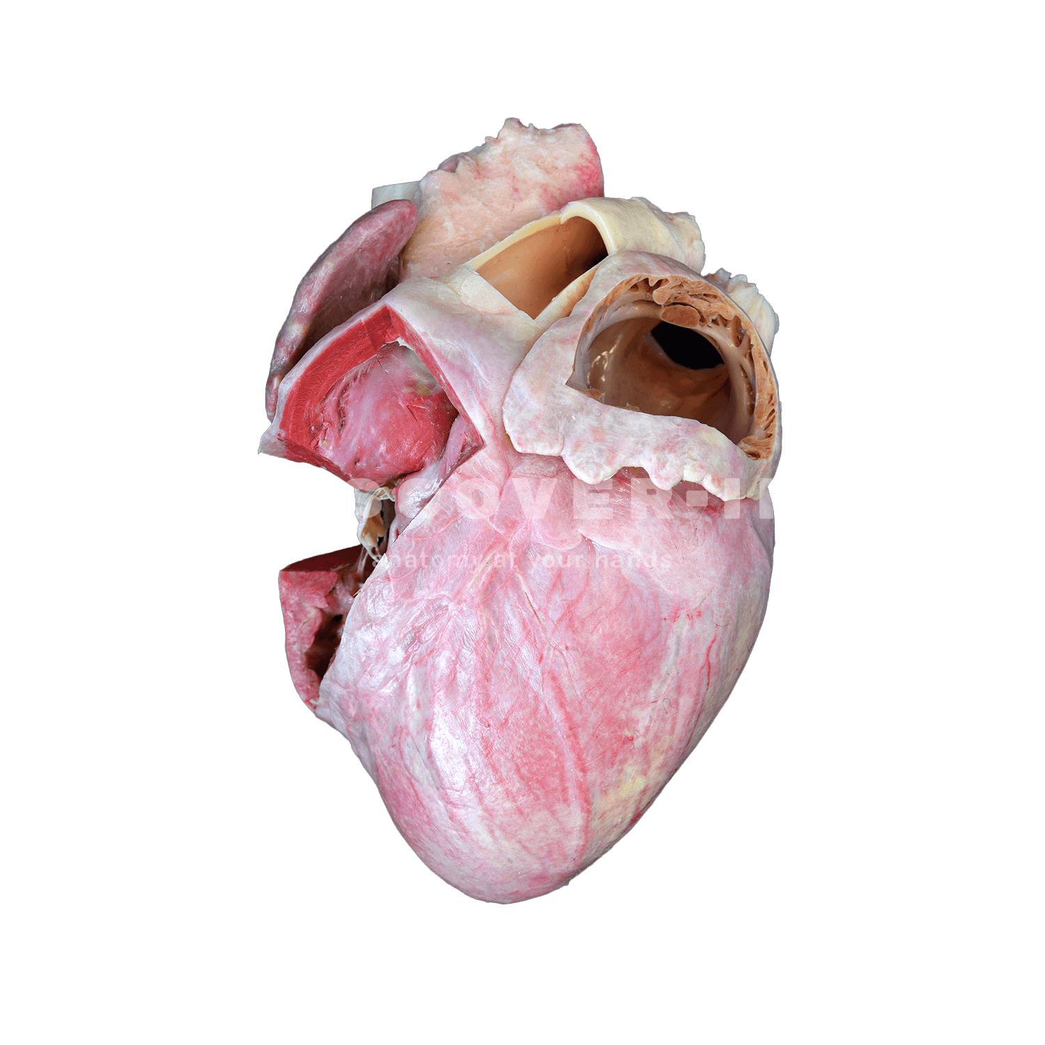
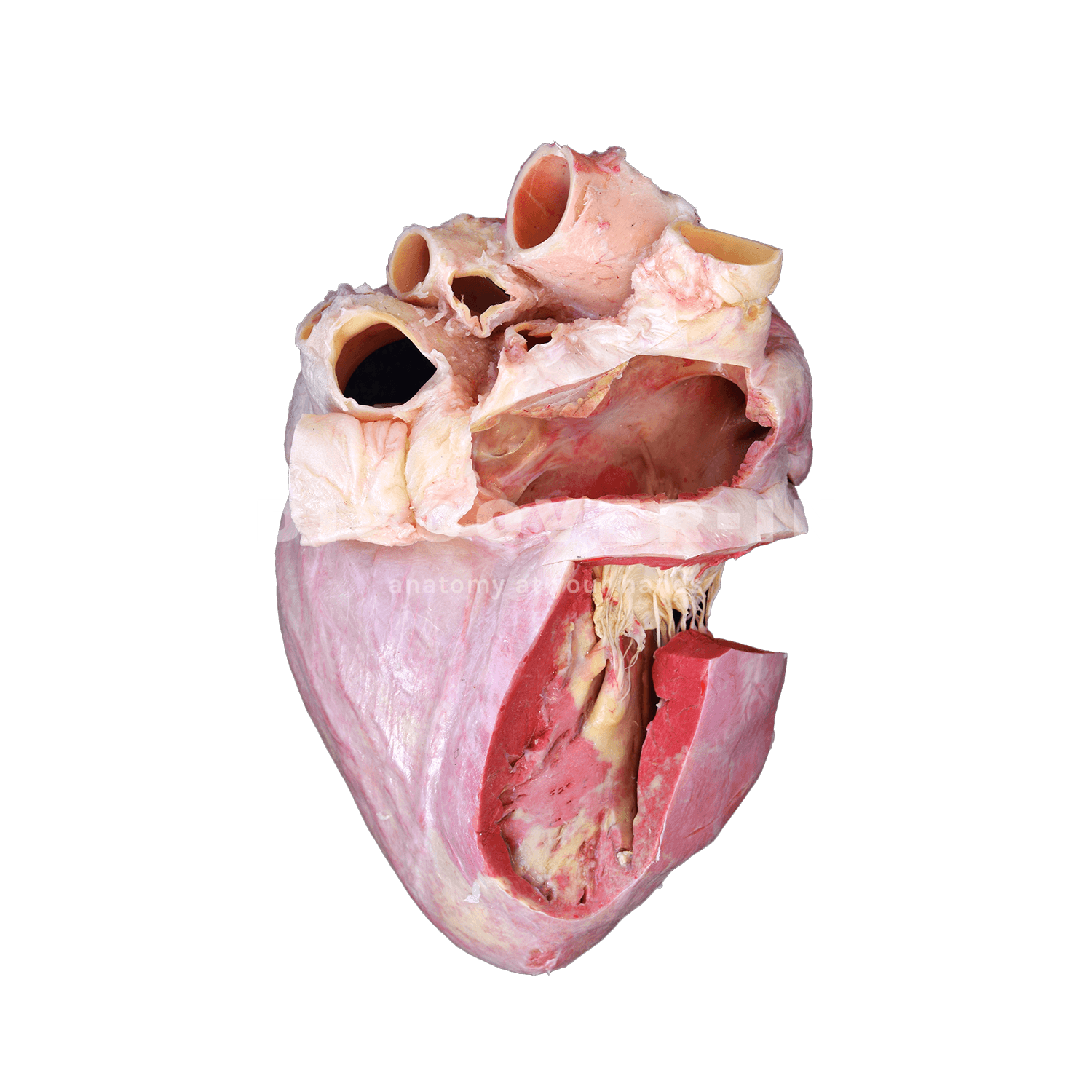
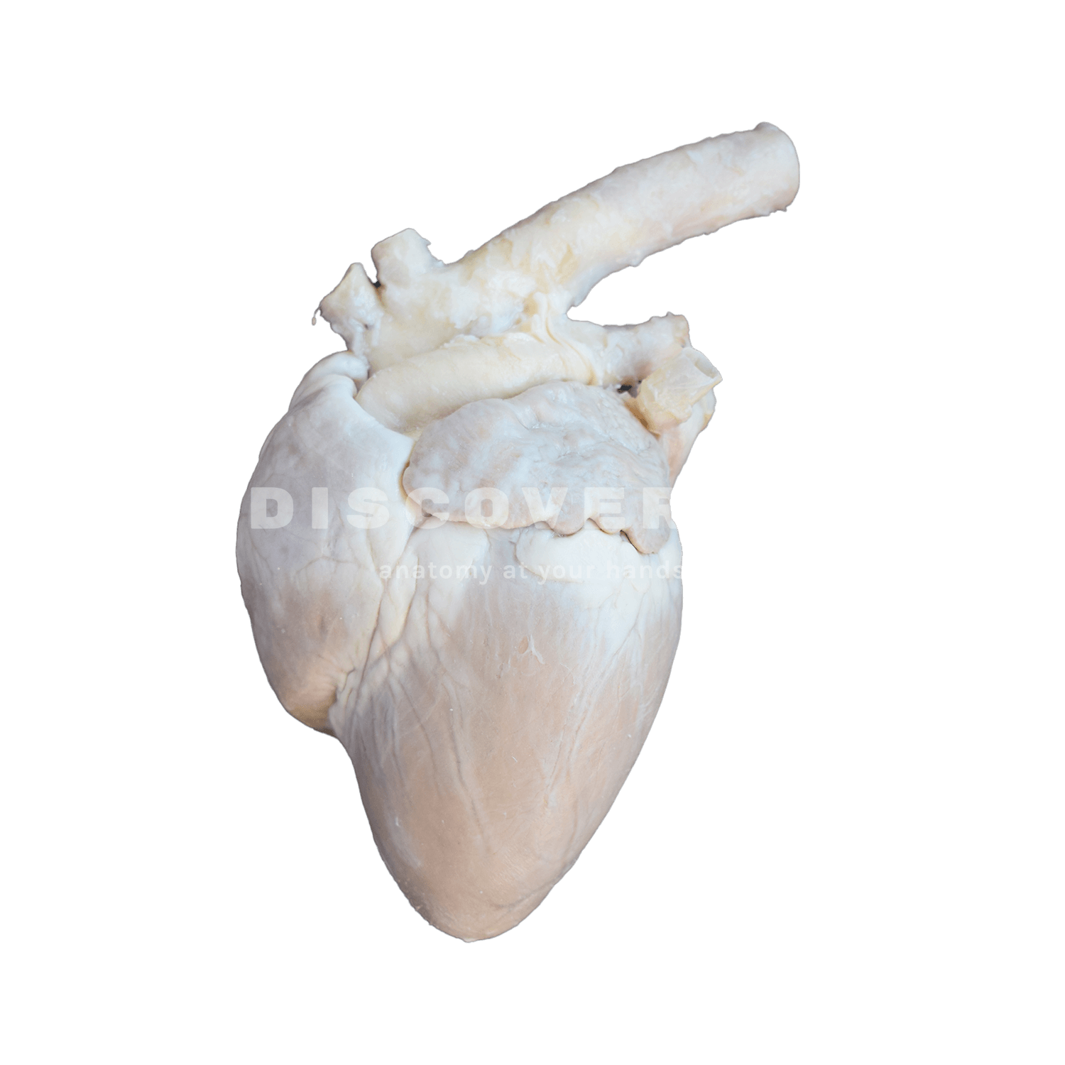
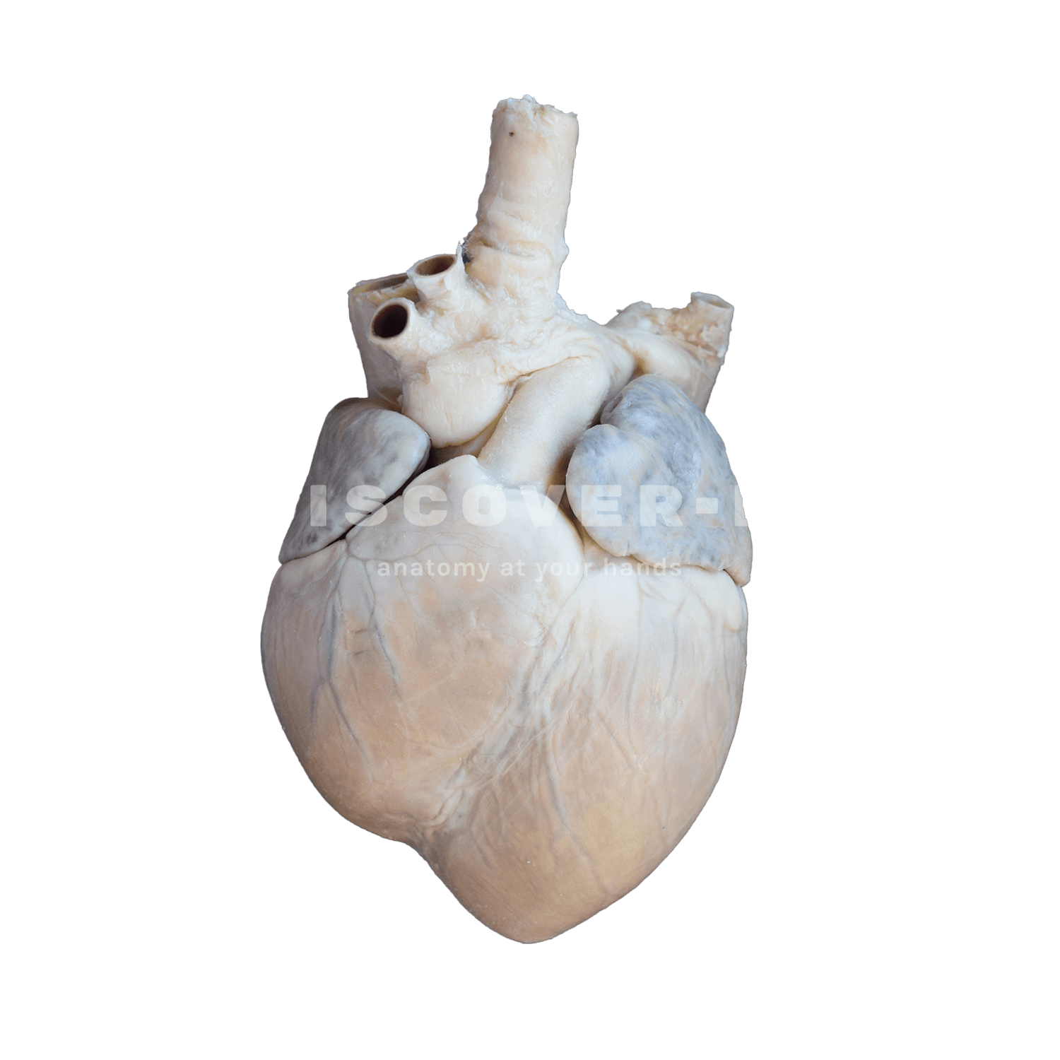
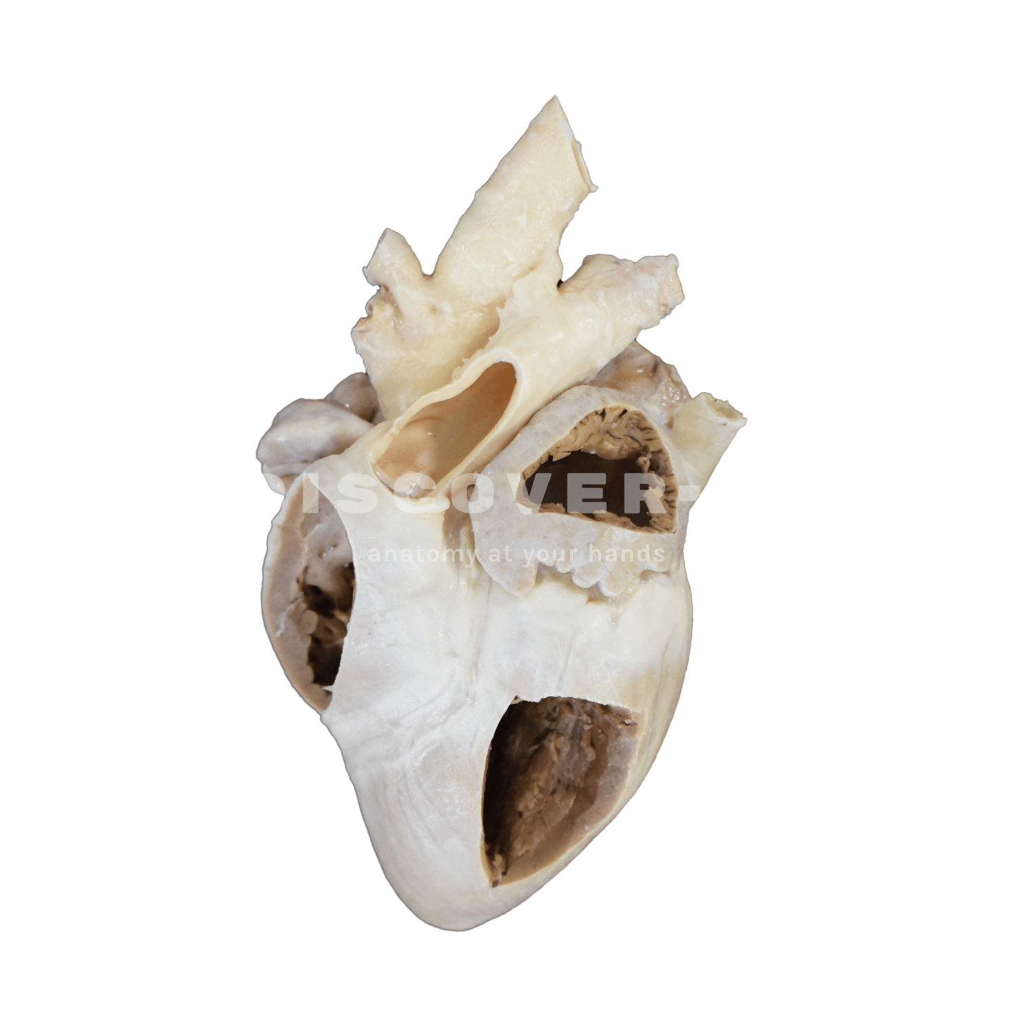
The complete heart shows the external morphology: apex, base, atria, ventriculi, grooves and great vessels. The dissected heart allows observation of the internal anatomy: cardiac cavities, valves, septa and muscular structures.
Comparing both helps relate the external surface with the internal organization and blood flow.
They are useful for teaching cardiovascular anatomy in domestic mammals.
The intact specimen helps identify external landmarks and the orientation of the heart.
The dissected specimen facilitates understanding of valvular function, ventricular wall thickness, and cardiac blood circulation.
Complete heart: apex, base, atria, ventricles, auricles, coronary grooves, coronary vessels, aorta, pulmonary trunk and major veins (cava veins).
Heart with dissection: ventricular cavities, atrial regions, interventricular septum, atrioventricular valves, semilunar valves, chordae tendineae, papillary muscles and trabeculae carneae.