
Ovine-Caprine flexible lungs
Ov-CpOpul
More images
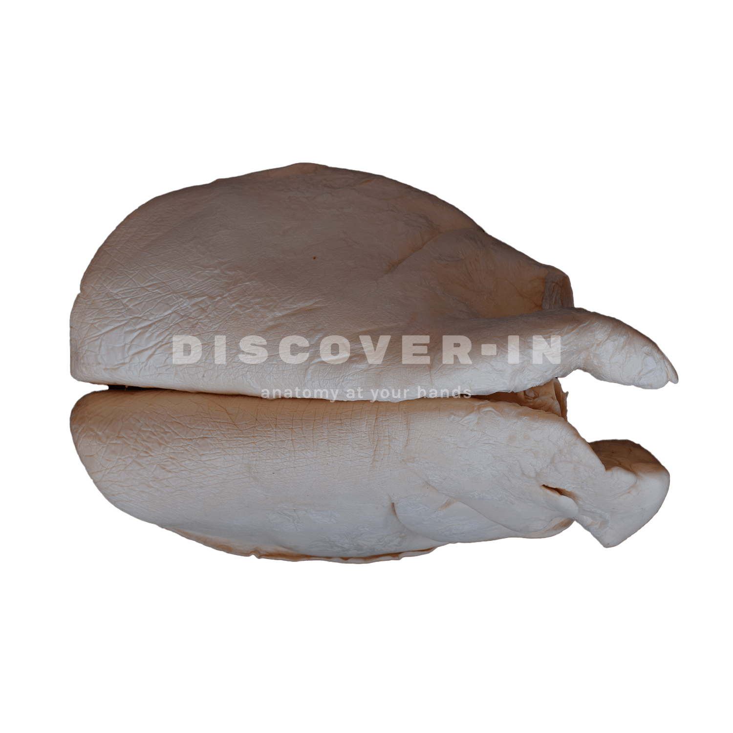
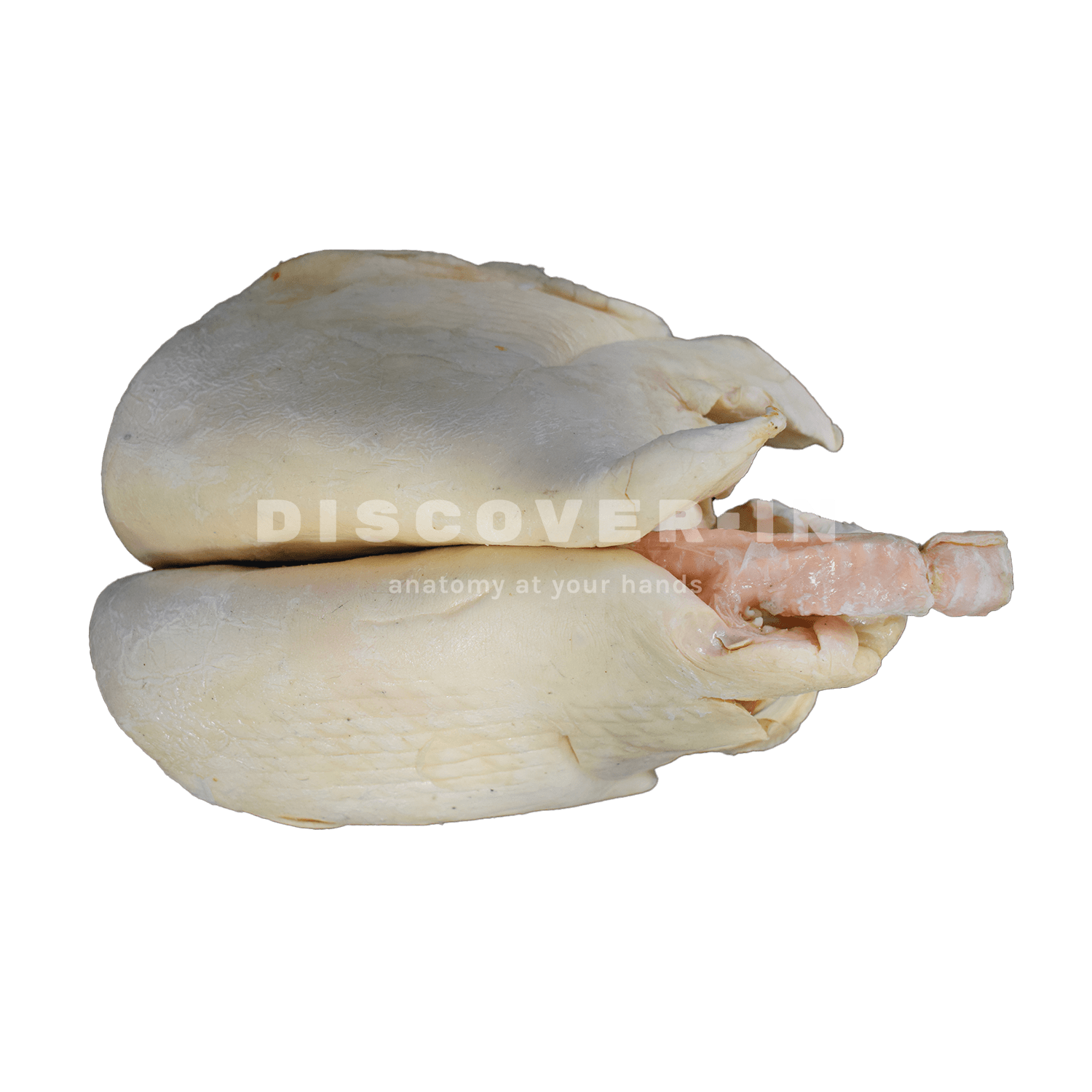
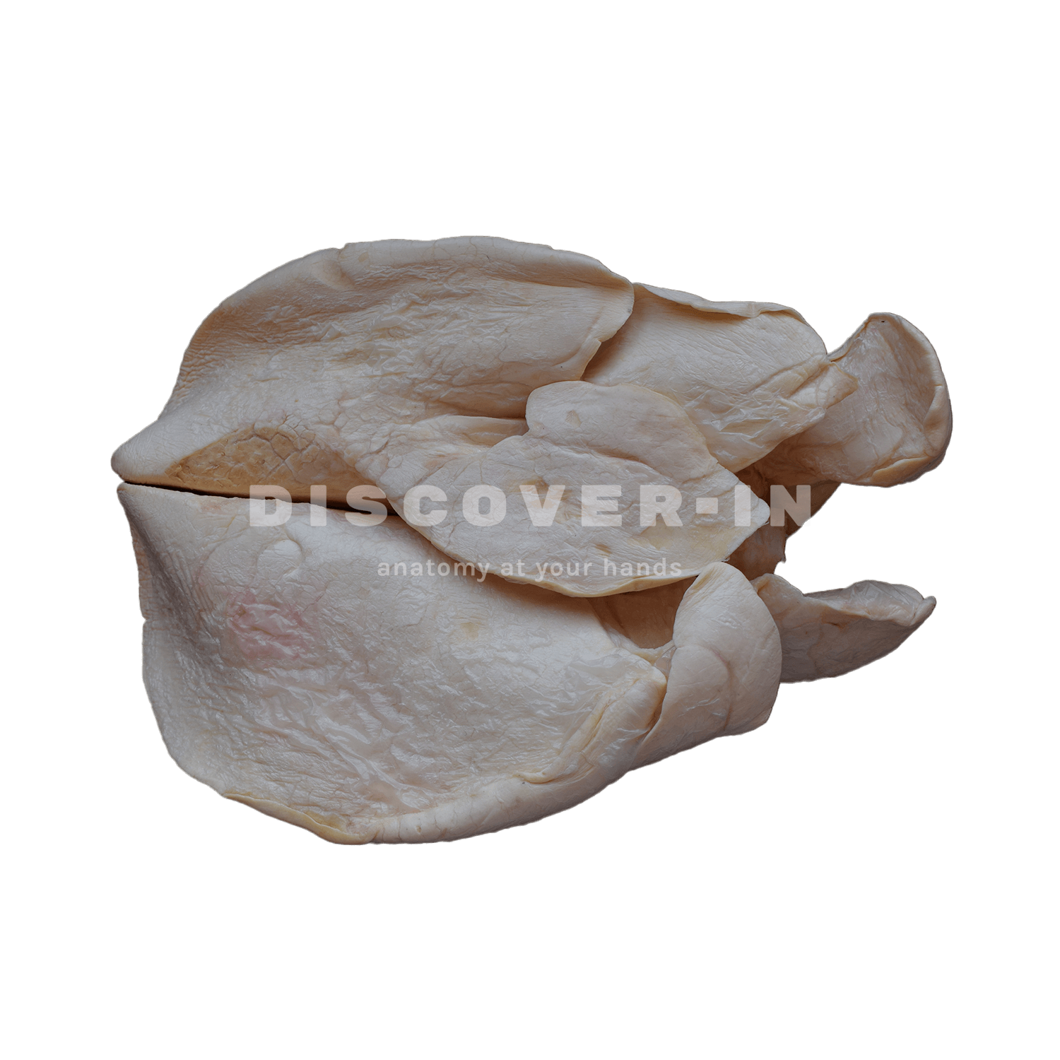
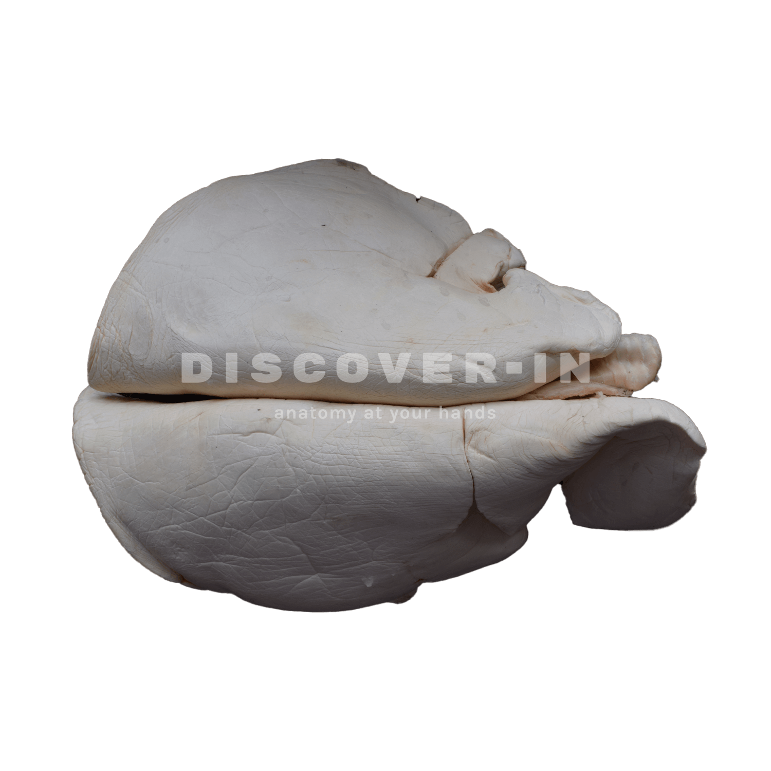
What is it in this specimen?
What can we learn from this specimen?
- Identify the carina and its continuation into the right and left main bronchi, linking airway anatomy with the pulmonary tissue.
- Recognise the visceral pleura and the general configuration of the lungs (lobes), which is useful for understanding their occupation within the thoracic cavity.
How can this specimen be used for teaching?
- Clinical topography: relate gross lung shape to auscultation and percussion zones in small ruminants (cranial vs caudal thorax).
- Airway procedures: use the visible trachea/bronchi to introduce endotracheal intubation and bronchoscopy orientation (airway branching at the hilus).
- Pathology discussion: teach where lesions such as consolidation, atelectasis or pleuritis would be expected and how pleural involvement changes clinical findings, without attributing disease to this specimen.