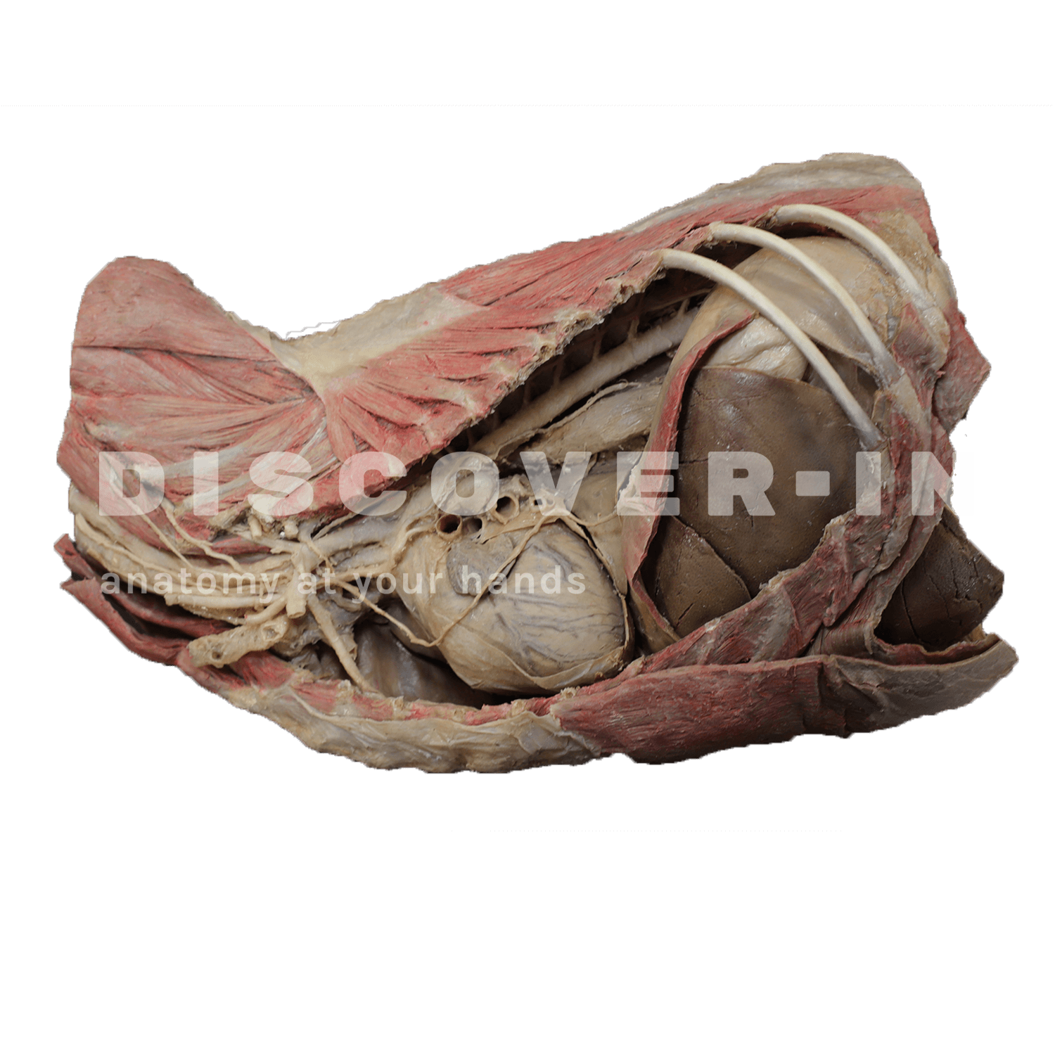
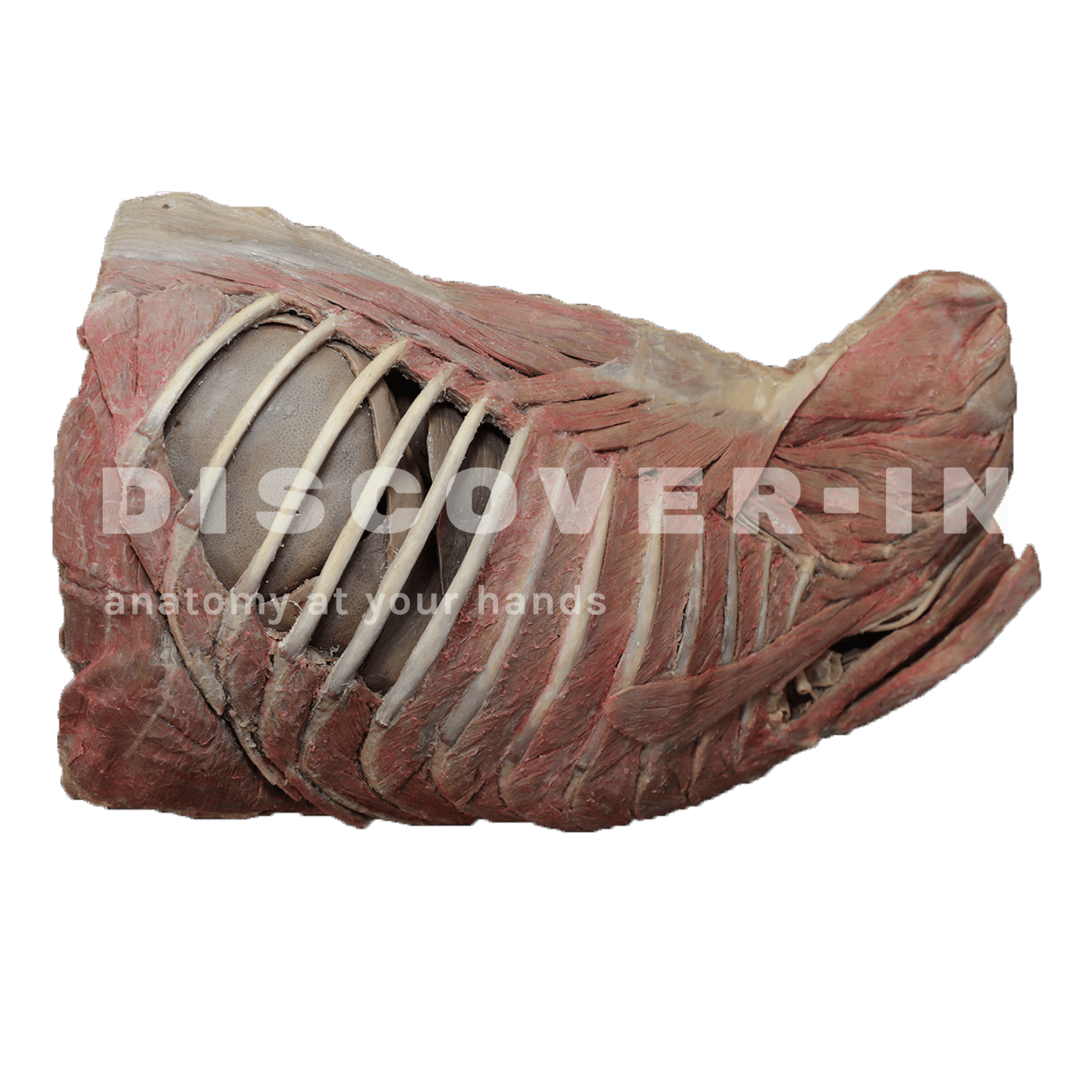
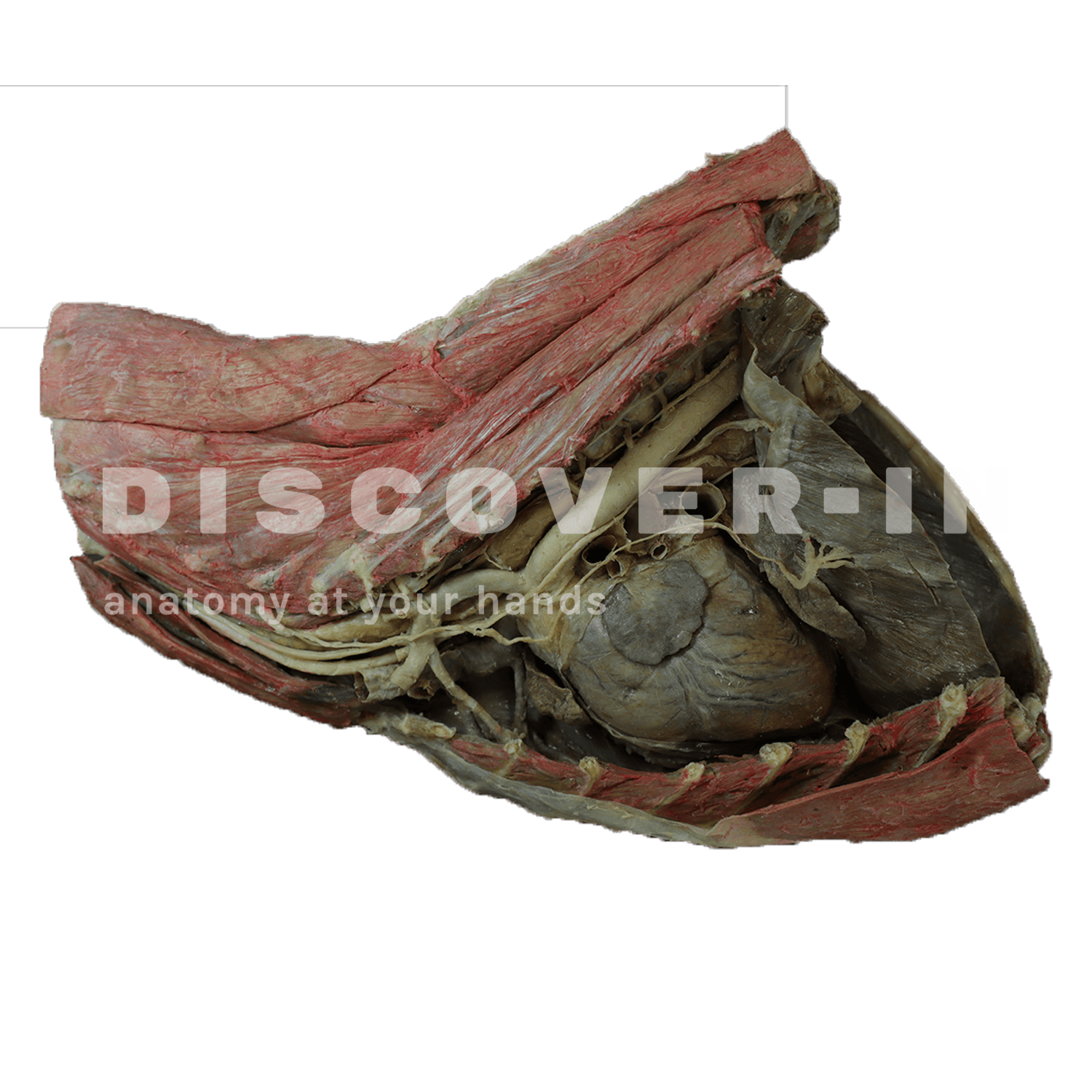
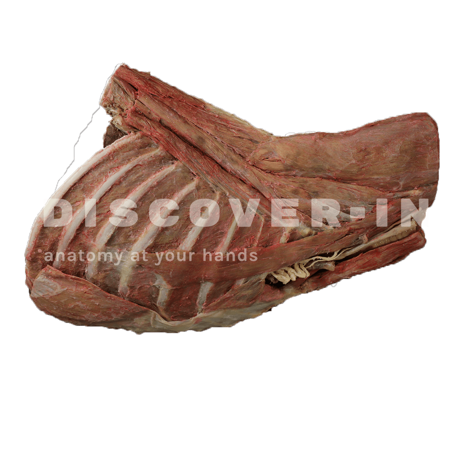
This specimen shows the topography of the heart after removal of the left lung during a dissection of the chest cavity and cranial abdomen of a dog approached from the left side. The cranial lobes and access of the permanent right lung in situ, serving as anatomical reference points. Within the cranial mediastinum, the brachiocephalic trunk and the left subclavian artery are identified as the main branches that arise from the aortic arch. The thoracic portion of the esophagus runs through the mediastinum in the craniocaudal direction. The diaphragm has been preserved to serve as a reference for retrodiaphragmatic organs such as the liver and stomach. The diaphragmatic pillars and their relationship with the aortic hiatus are preserved, as are the initial visceral branches of the abdominal aorta, especially the celiac trunk and the cranial mesenteric artery. Visible son the interventricular and coronary grooves. A portion of the right atrial wall has been removed to expose the right atrioventricular valve ( tricuspid ), including the tendon cords and papillary muscles. In addition, the septomarginal trabecula ( moderating band ) is observed within the right ventricle, as well as its trajectory from the ventricle to the pulmonary trunk. The left atrial wall is also open to visualize its light.