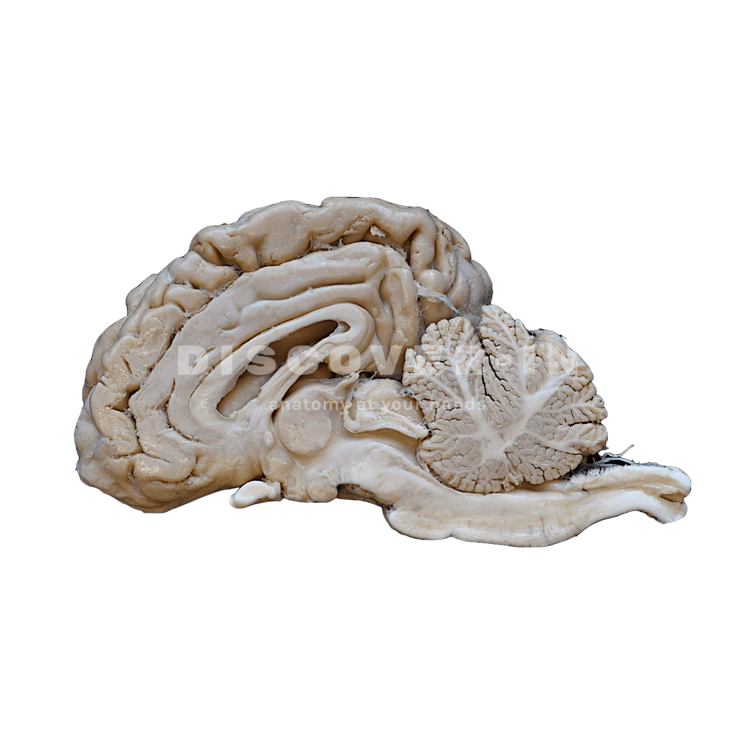
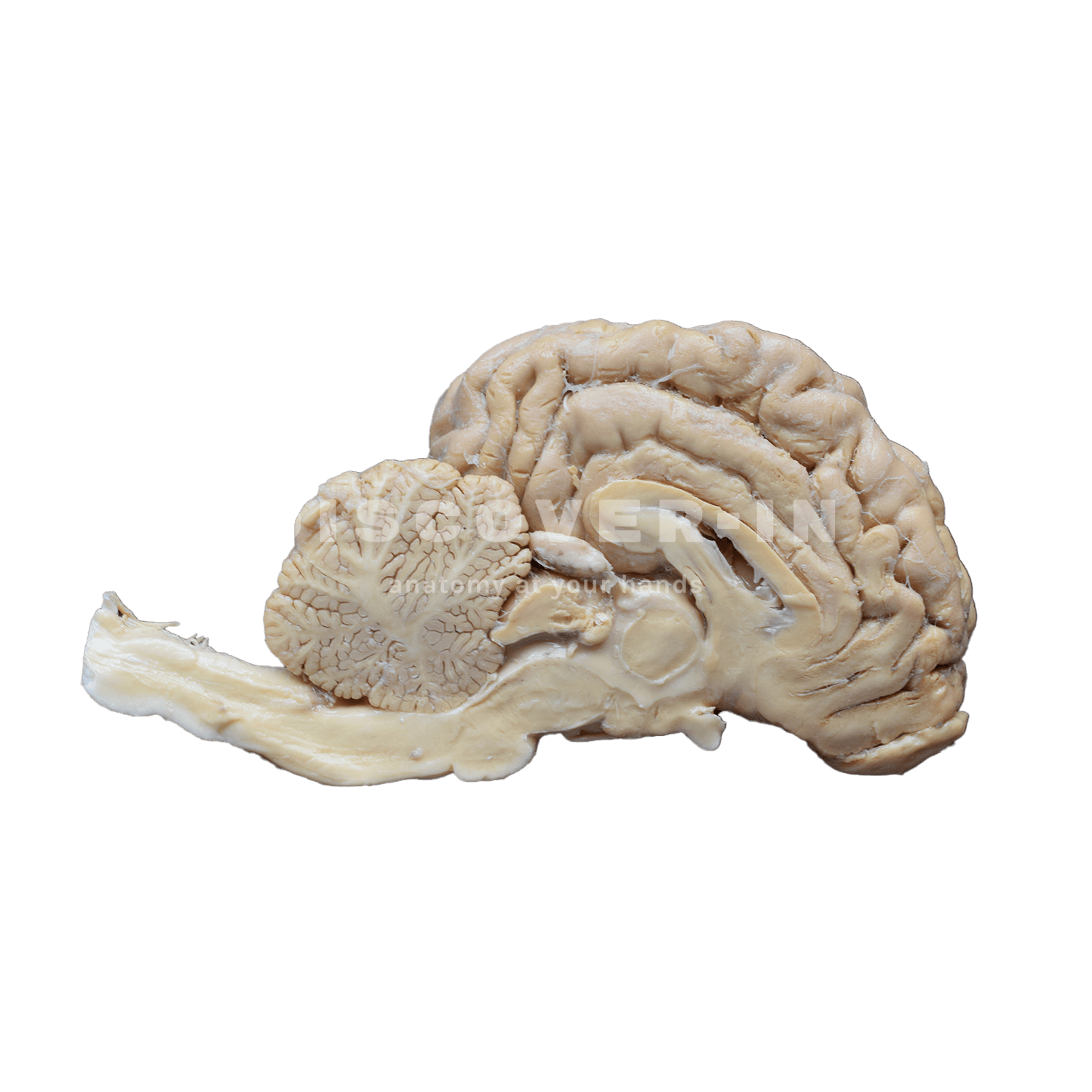
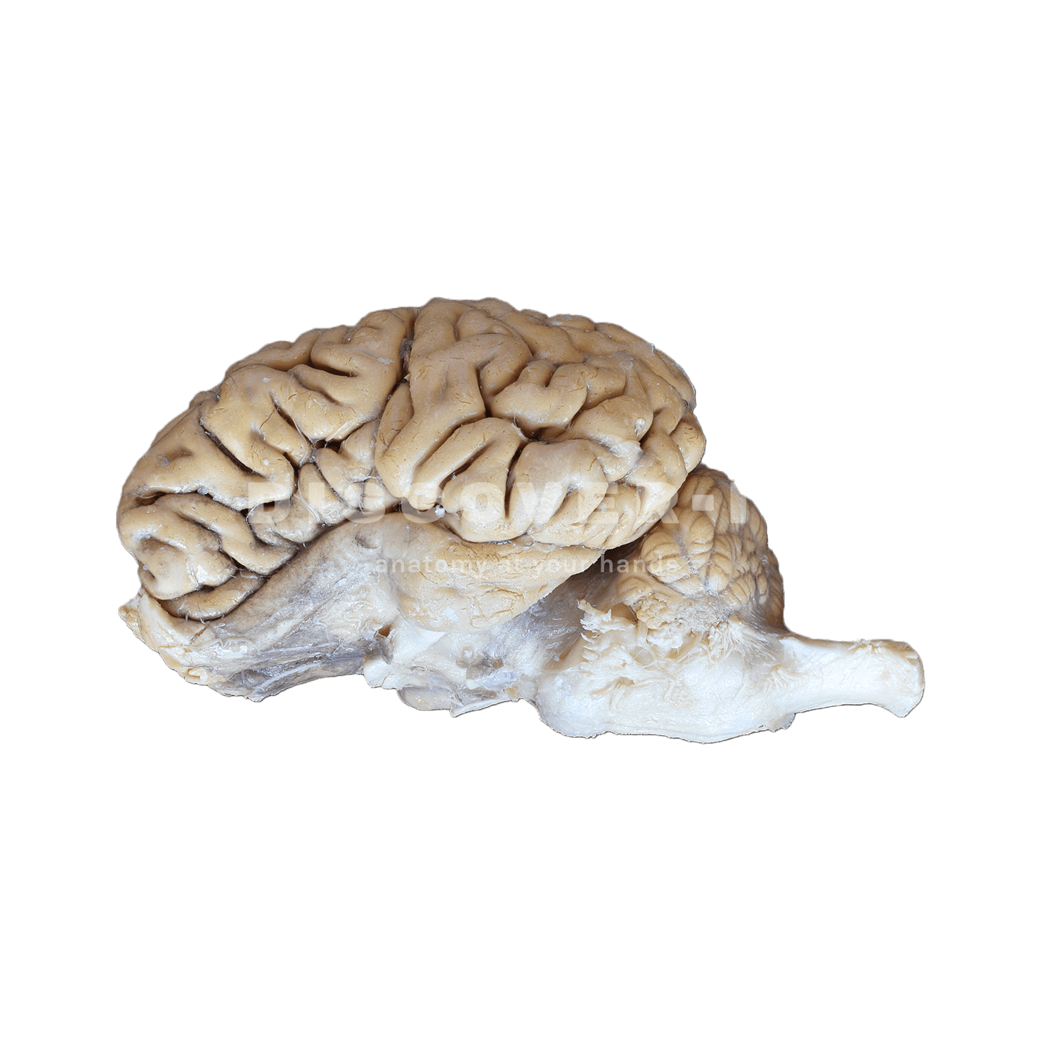
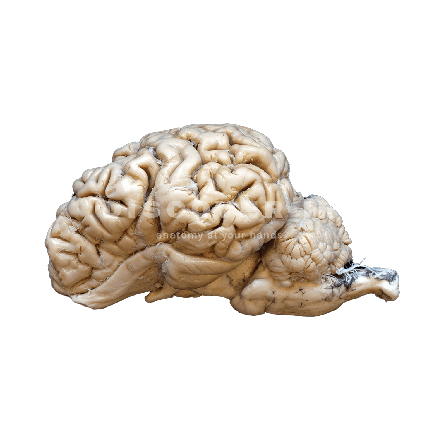
This specimen correspond to a plastinated hemiencephalon of a bovine, shown in lateral view (external surface) and in medial view after a sagittal section. In the lateral aspect, the cerebral hemisphere exhibits a well-developed gyrencephalic cortex with prominent gyri and sulci. Caudally, the cerebellum is visible with clearly defined cerebellar hemispheres and vermis, and ventrally the brainstem can be identified, including the mesencephalon, pons, and medulla oblongata in continuity with the spinal cord. In the medial (sagittal) section, the corpus callosum is distinctly observed arching over the lateral ventricle, the fornix ventral to it. The diencephalon is represented by the thalamic region and hypothalamic area, and the third ventricle is identifiable in the midline. The cerebellar arbor vitae is clearly visible within the cerebellar parenchyma, and the fourth ventricle lies between the cerebellum and the brainstem.
- To recognize the topographical relationships between telencephalon, diencephalon, cerebellum, and brainstem in a large domestic species.
- To identify major commissural and ventricular structures in sagittal section, particularly the corpus callosum and third ventricle.
- To understand the organization of the cerebellum, including the arbor vitae and its relationship to the fourth ventricle.
- To train students in correlating external brain morphology with internal sagittal anatomy, reinforcing three-dimensional understanding.
- To demonstrate clinically relevant regions such as the brainstem and cerebellum in relation to neurological deficits (e.g., ataxia, cranial nerve dysfunction).
- To support interpretation of mid-sagittal MRI images by direct anatomical comparison with real specimens.