Bovine brain serial sagittal sections 3-4 mm thick (6-8 sections)
BoOencP40s
More images
What is it in this specimen?
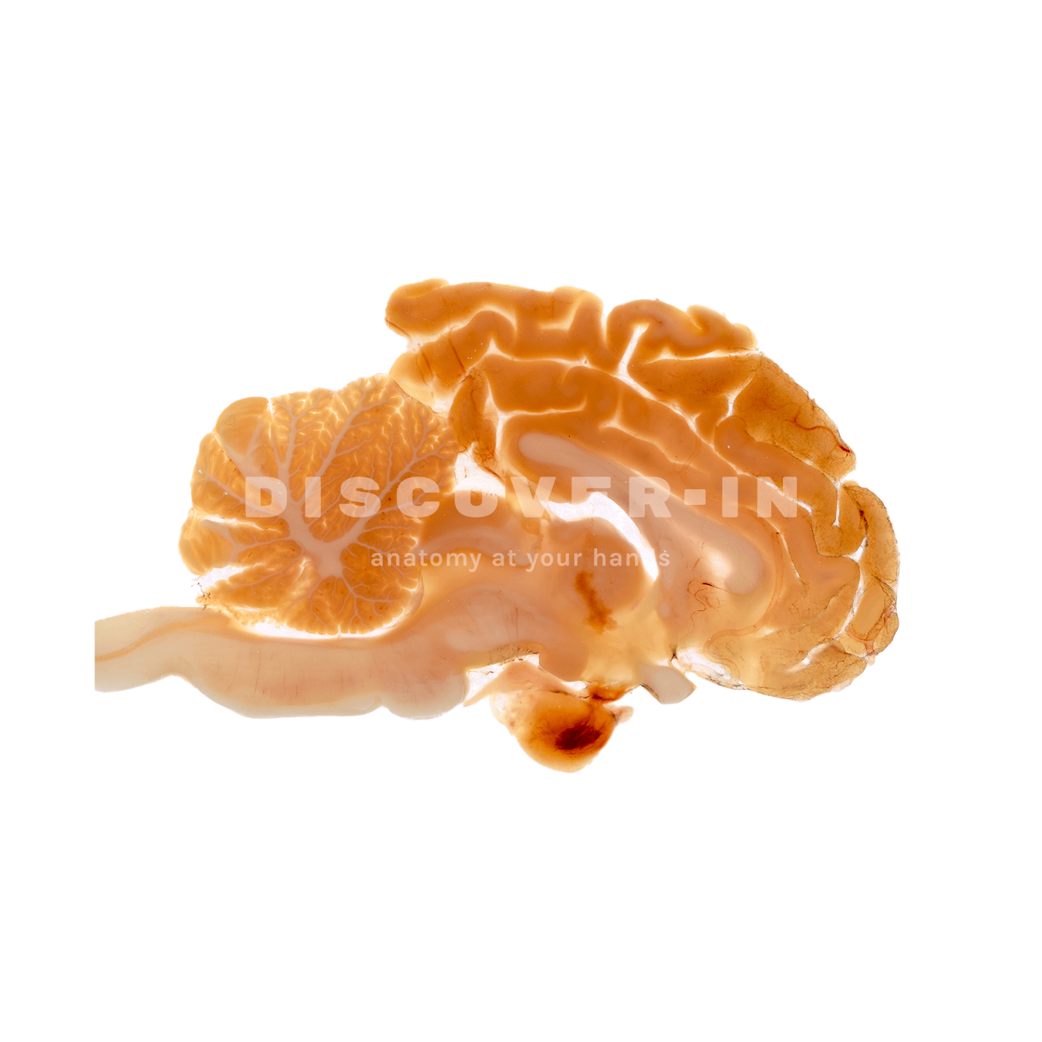
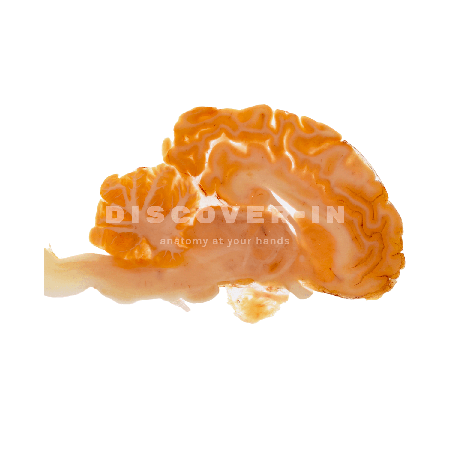
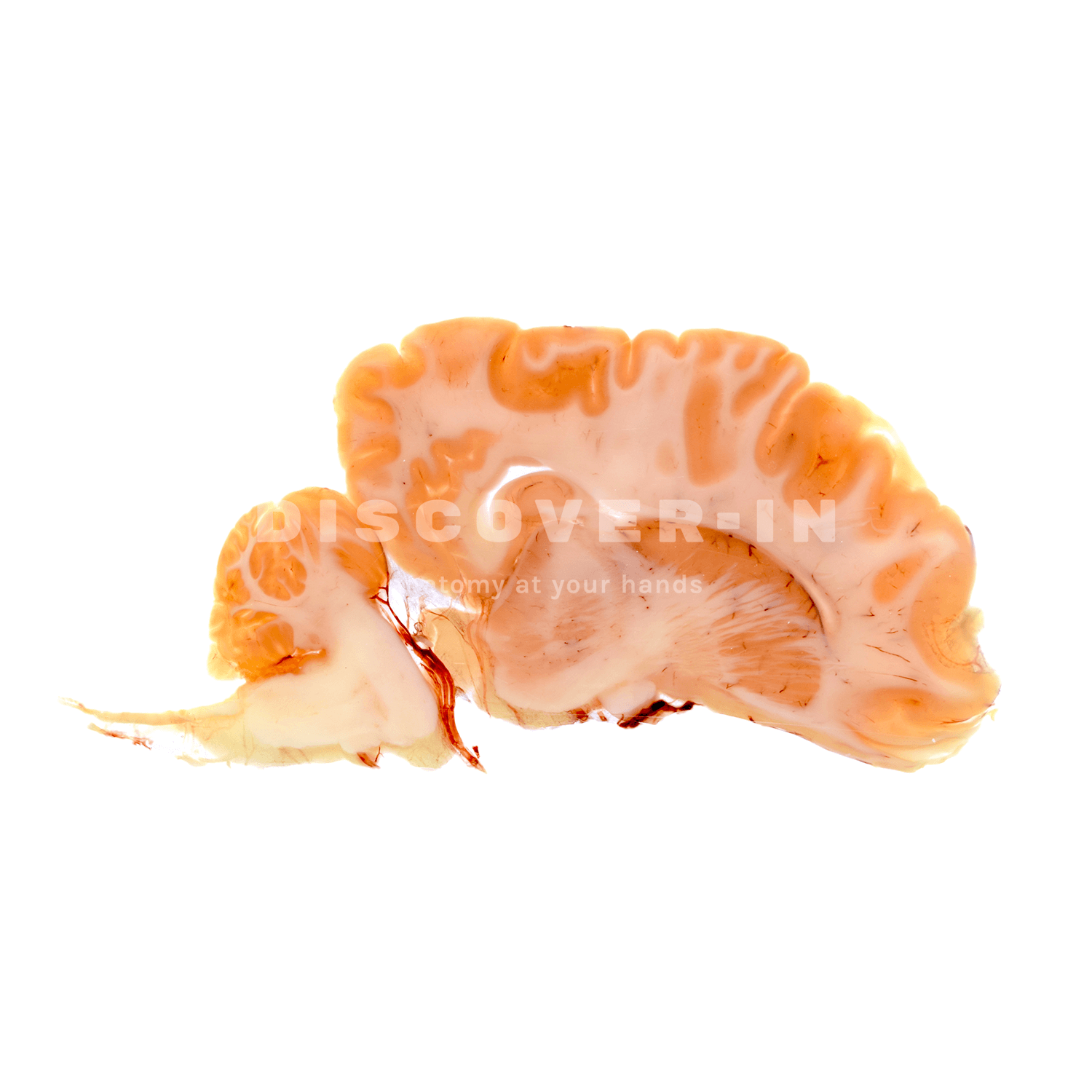
This is a series of sagittal sections of a bovine brain plastinated using the P40 technique, showing the medial surface of a cerebral hemisphere with its cortical gyri and sulci, as well as the underlying cerebral white matter.
Caudodorsally, the cerebellum is visible, with its characteristic folia and internal white matter, the arbor vitae. In a ventral and caudal position, the brainstem can be identified, allowing the continuity between the diencephalic region and the midbrain/pons–medulla oblongata complex to be appreciated in the same plane.
What can we learn from this specimen?
- Correlate gray vs white matter organization: cortical mantle (gray matter) surrounding deeper cerebral white matter, and the cerebellar cortex surrounding the arbor vitae.
- Understand the topographic relationships between cerebrum, cerebellum, and brainstem in a sagittal plane (rostrocaudal and dorsoventral orientation).
How can this specimen be used for teaching?
- As a reference for sectional neuroanatomy, linking gross dissection to sagittal imaging planes used in CT/MRI interpretation in large-animal practice.
- For clinical localization exercises: use the specimen to discuss how lesions affecting cerebellum vs brainstem vs cerebral hemisphere can produce different neurological deficits (e.g., ataxia vs cranial nerve/brainstem signs vs altered mentation).
- To teach neuroanatomical pathways in context (e.g. cerebellar coordination circuits and brainstem integration) by tracing structures in a continious, undistorted sagittal view.