Swine Stomach
SuOest
More images
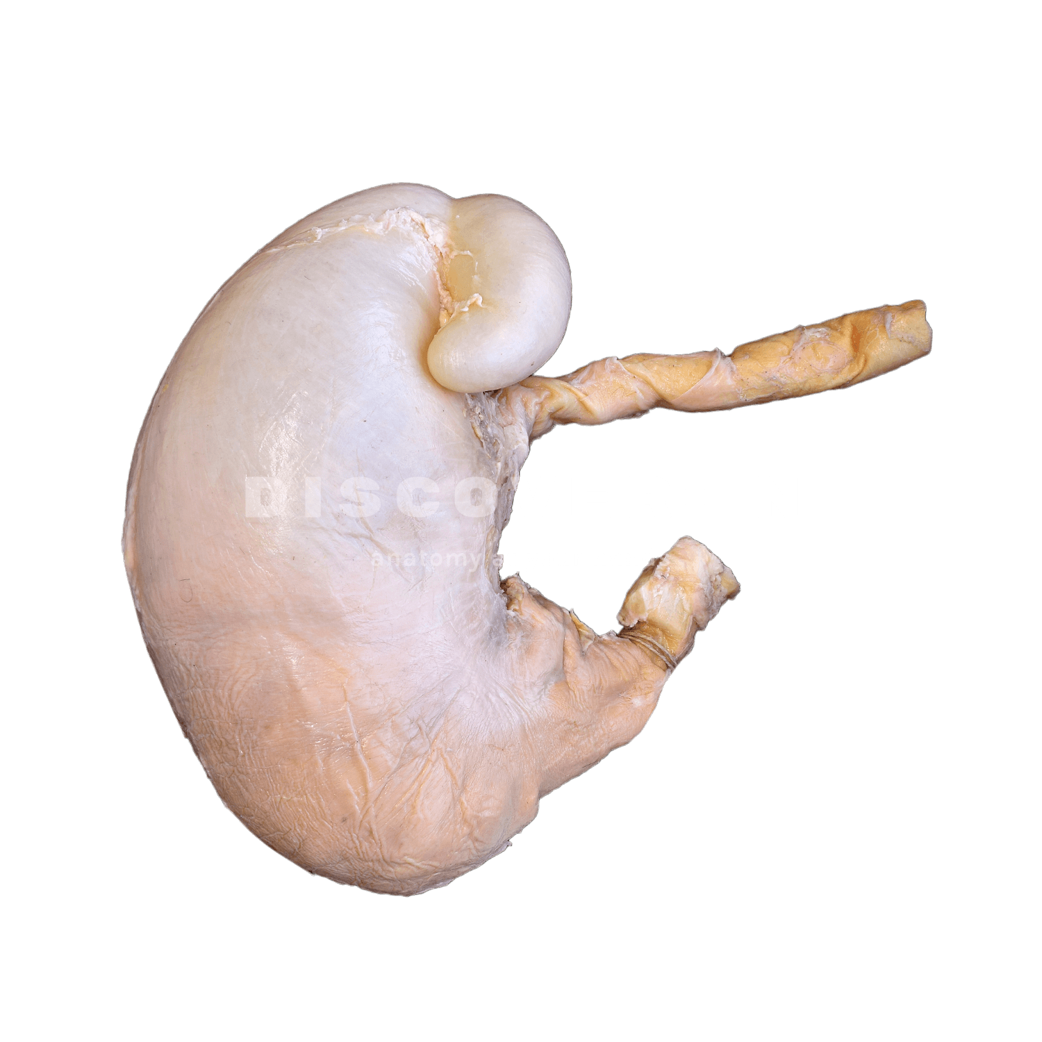
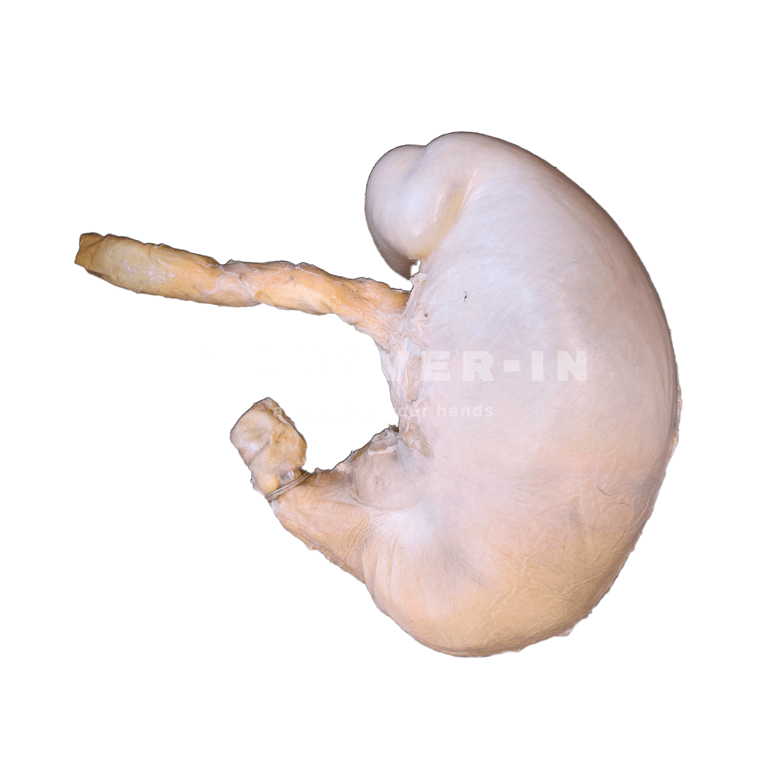
What is it in this specimen?
It is a plastinated swine stomach.
The greater curvature is clearly visible as the dominant convex border, and in the proximal region a well-defined gastric diverticulum (diverticulum ventriculi), characteristic of the pig, can be seen adjacent to the fundus.
The stomach narrows towards the pyloric portion, continuing into a short segment of the proximal duodenum.
What can we learn from this specimen?
- Species-specific landmark: the gastric diverticulum is a key porcine feature and helps orient the fundic region relative to the rest of the stomach.
- Regional anatomy: appreciate the gross separation of fundus/body versus pyloric region, and how the stomach narrows approaching the pylorus and duodenum.
- External topographical orientation: use the greater curvature as a guide to the overall morphology and the site of omental attachment
How can this specimen be used for teaching?
- Clinical orientation drills: have students trace the route esophagus → cardia → fundus/body → pylorus → duodenum, reinforcing where obstructions, ulcers, or foreign bodies would be expected clinically.
- Comparative anatomy: contrast the porcine stomach (including the diverticulum ventriculi) with dog/ruminant stomachs to train rapid species recognition in imaging and surgery.
- Surgical landmarks: use the external morphology to discuss approaches to the pyloric outflow and proximal duodenum, emphasizing safe identification of regions before any enterotomy/gastrotomy planning.