Canine stifle joint
CaProd
More images
What is it in this specimen?
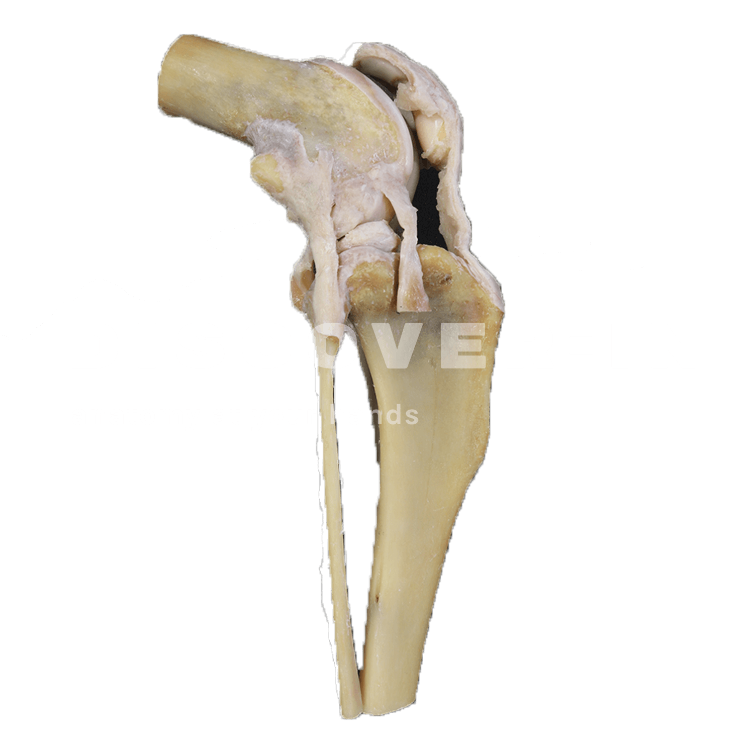
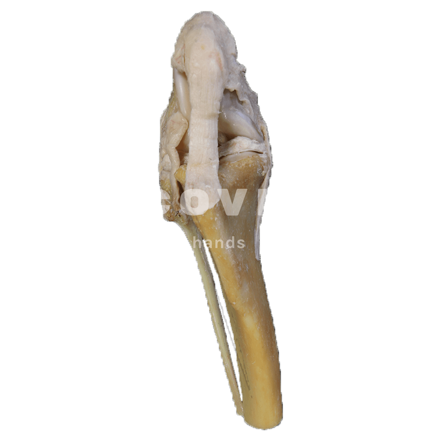
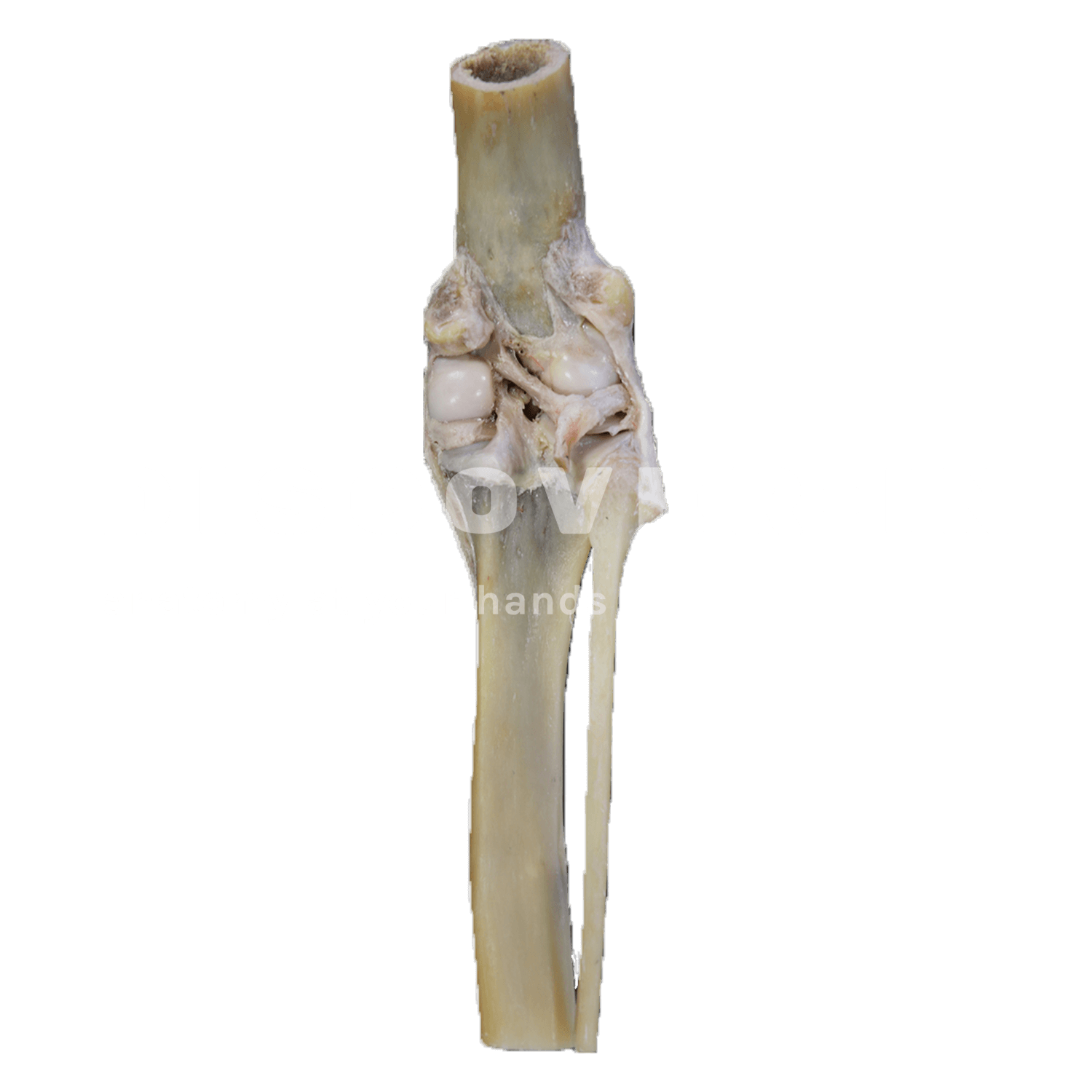
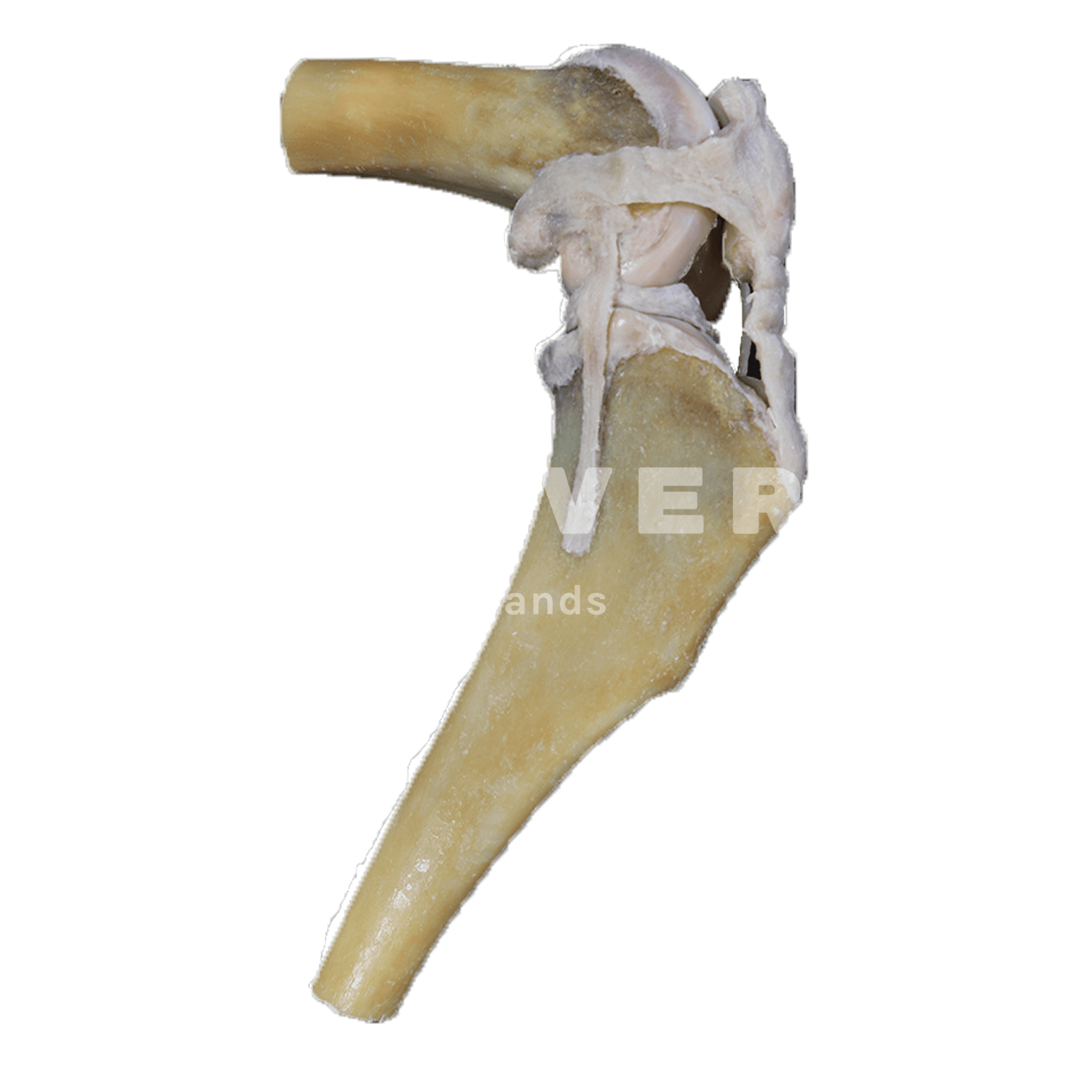
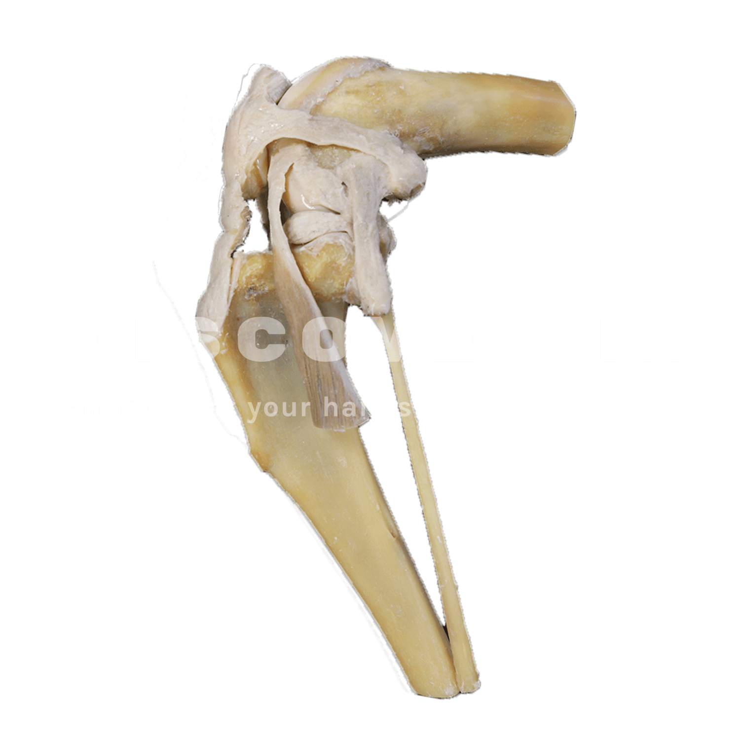
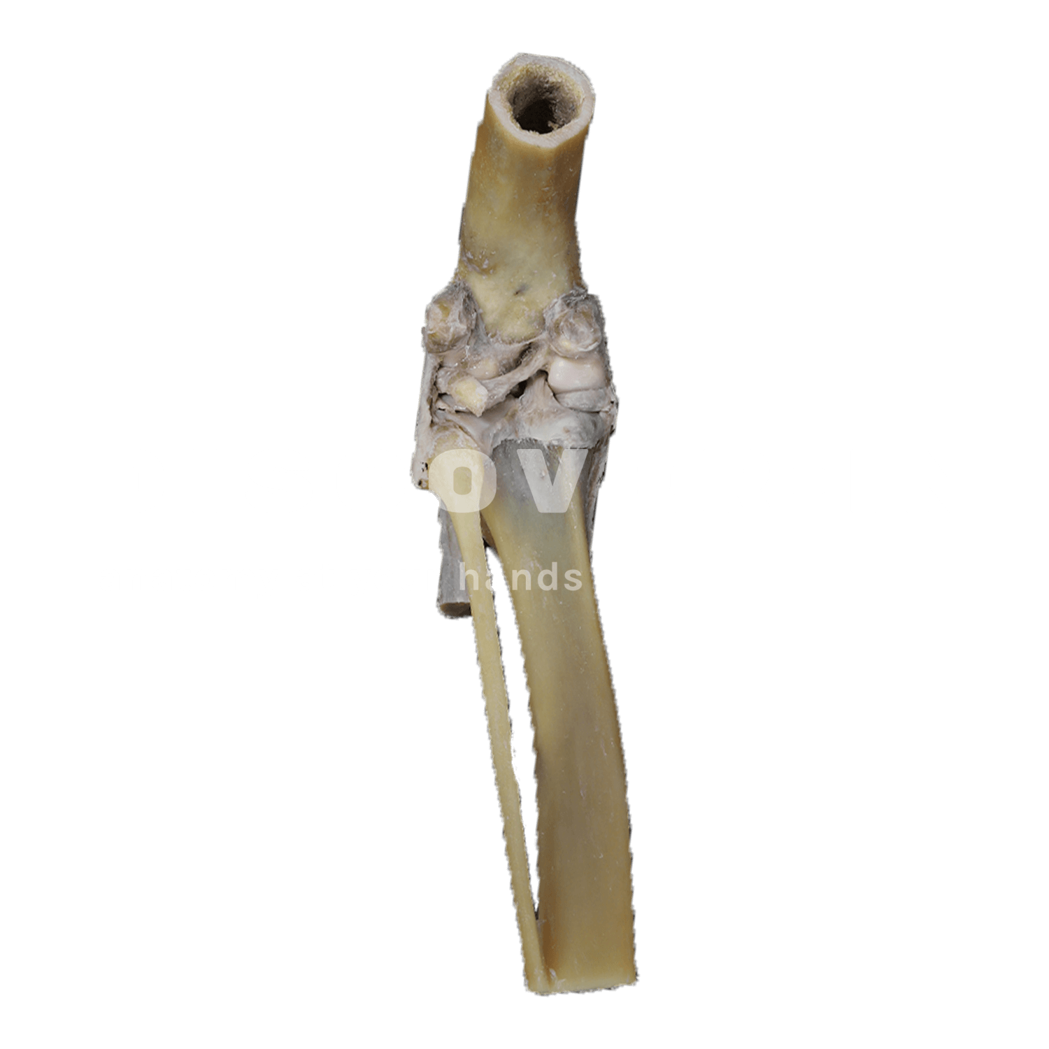
This plastinated specimen shows the canine stifle joint (knee), a synovial hinge joint formed by the distal femur, proximal tibia and fibula, patella and gastrocnemius sesamoid bones. It highlights the stabilizing soft tissues that guide and restrict motion, including the cranial and caudal cruciate ligaments, the medial and lateral collateral ligaments, the medial and lateral menisci, and the patellar ligament as part of the extensor mechanism.
What we can learn from this specimen?
- Identify the main osseous components and relate their articular surfaces to stifle flexion–extension mechanics.
- Understand how the cranial cruciate ligament and menisci contribute to cranial–caudal stability and load distribution.
- Link palpation-based assessment to anatomy by visualizing how cranial translation of the tibia increases when cranial cruciate integrity is compromised (cranial drawer concept).
How can be this specimen used for teaching?
- Use it for practical demonstrations of joint stability: correlate the “cranial drawer test” with the anatomical position and function of the cruciate ligaments.
- Support orthopedic case discussions (e.g., suspected cranial cruciate ligament rupture) by mapping clinical signs to specific intra-articular structures.
- Reinforce pre-surgical orientation by tracing likely surgical landmarks and discussing how meniscal injury may accompany cruciate pathology.