

If you have access to our plastinated specimens, you will be able to deepen your anatomical knowledge in a practical and hands-on way.These specimens will enable you to:
• Precisely identify key anatomical structures.
• Understand anatomical reality in detail.
• Reinforce theoretical learning.
• Strengthen the acquisition of practical and clinical skills.
These advantages make Discover-IN specimens an innovative resource for students, educators, and professionals seeking comprehensive, effective, long-lasting, and biologically safe training in veterinary anatomy.
Discover-IN provides real plastinated specimens designed specifically for practical, safe, and long-lasting veterinary anatomy education and training. These materials are ideal for veterinary faculties, anatomy laboratories, and continuing education centers that require high-quality anatomical resources.
The catalog includes a wide range of species and anatomical systems, offering specimens that support hands-on understanding—from isolated organs to complex dissections, body sections or organ blocks. Each piece is unique thanks to the plastination process.
This resource adds significant value to veterinary training by enhancing learning through direct experience and complementing theoretical knowledge with real, easy-to-handle materials.
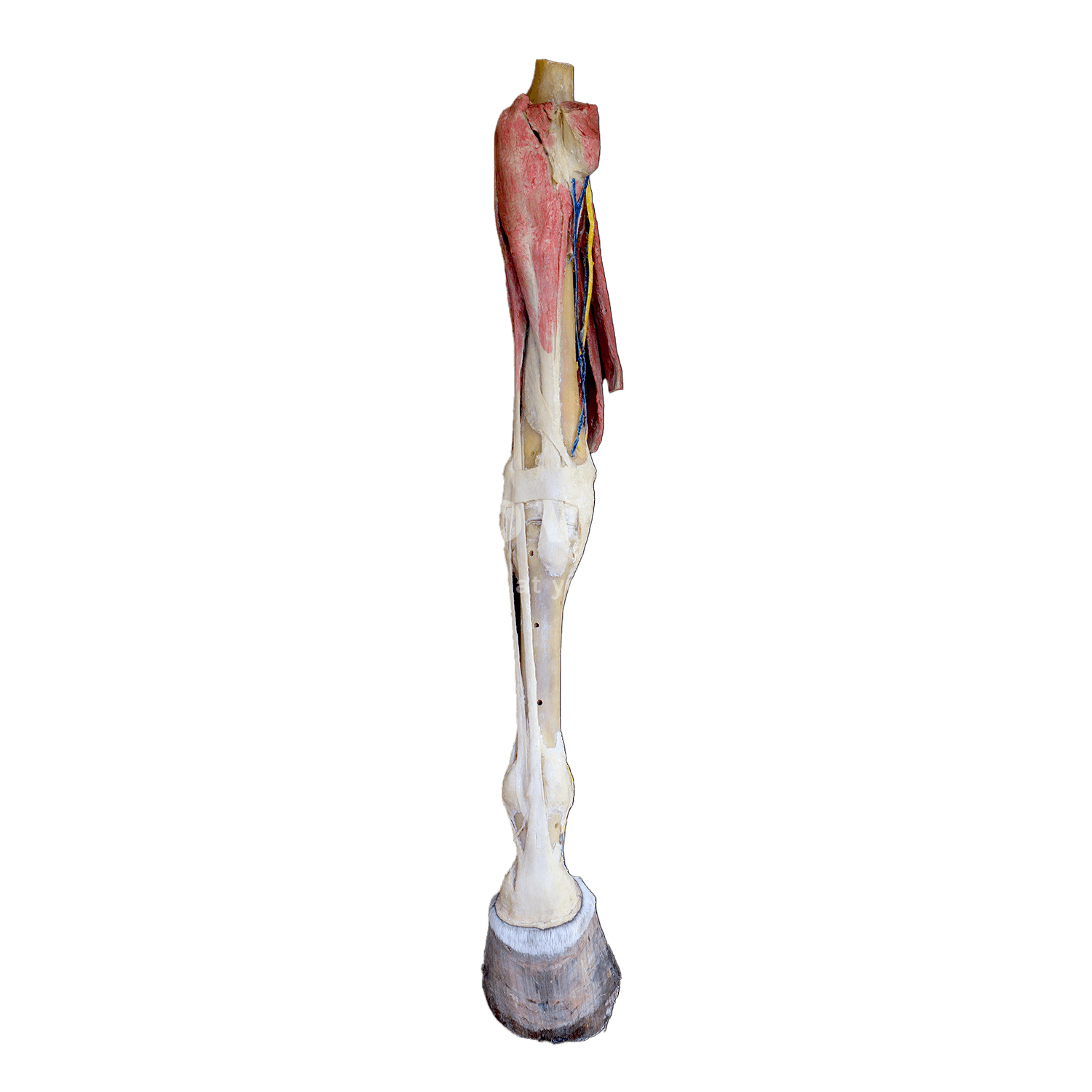
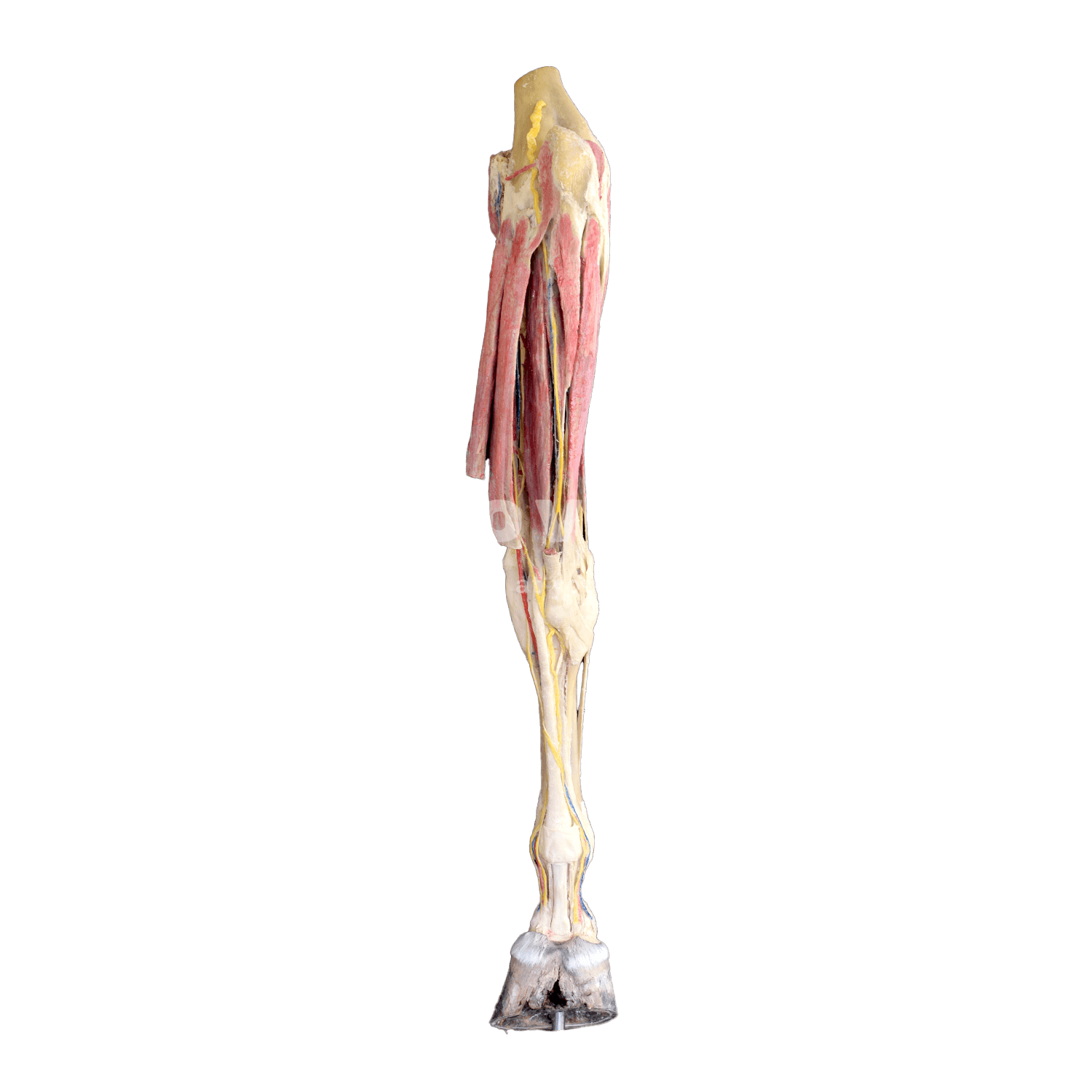
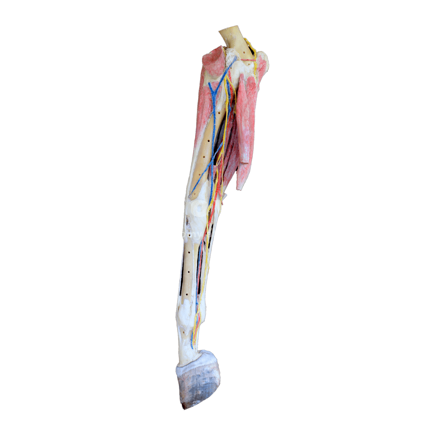
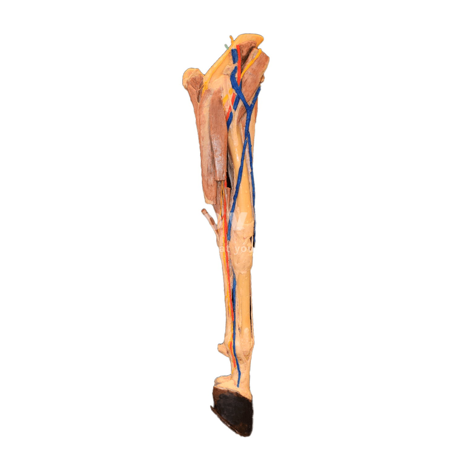
The equine forelimb is one of the most specialized and biomechanically sophisticated structures in the animal kingdom. It plays a critical role in locomotion, impact absorption, weight support, and athletic performance. A comprehensive understanding of its anatomy is crucial for accurately diagnosing lameness, designing effective rehabilitation programs, and performing precise surgical interventions. This anatomical expertise is also indispensable for the accurate interpretation of diagnostic imaging, including radiography, ultrasonography, computed tomography (CT), and magnetic resonance imaging (MRI).
1. Skeletal Structure of the Forelimb
The skeletal framework of the equine forelimb combines strength, elasticity, and precision:
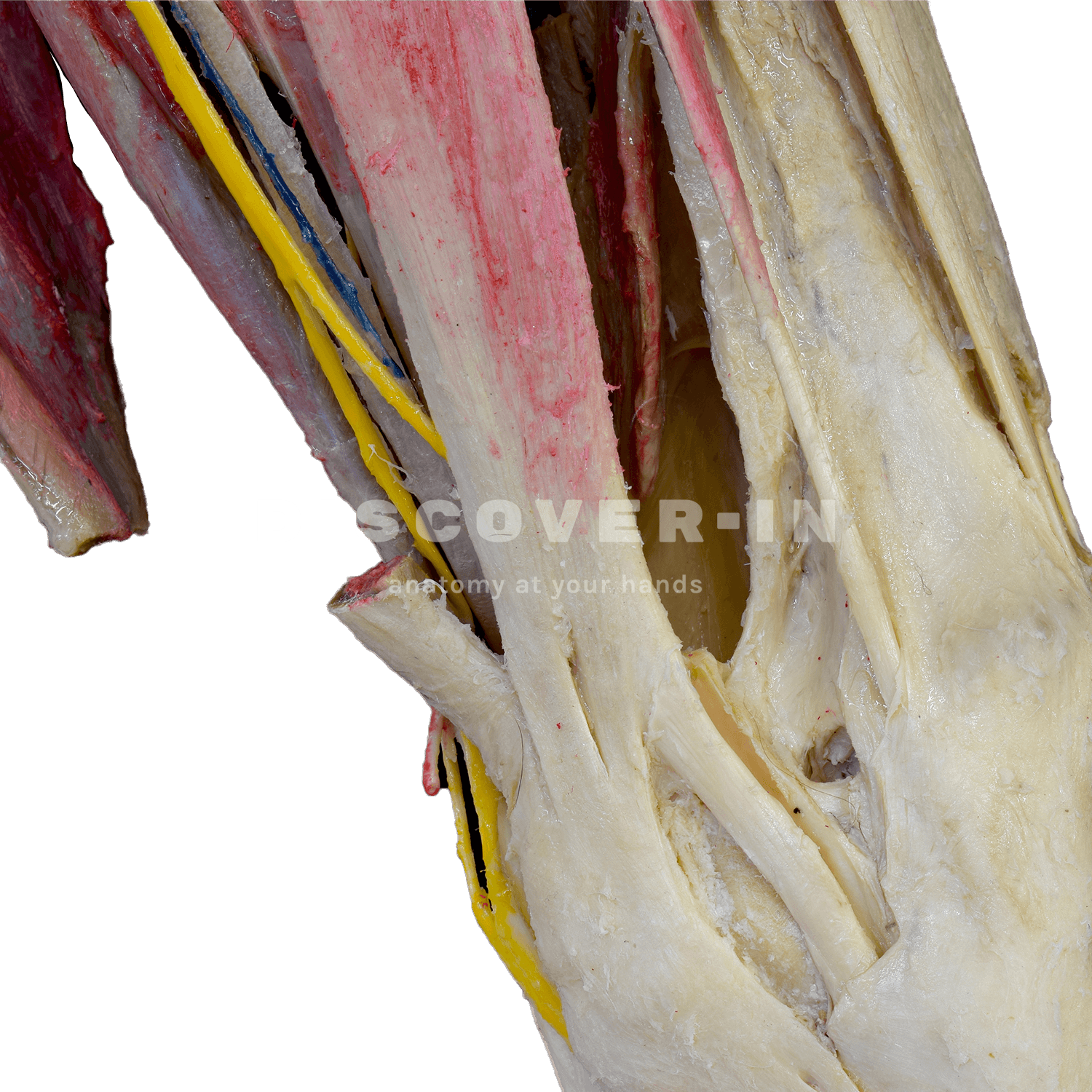
- Scapula (shoulder blade): Connected to the thorax only by muscles — not by a bony joint — creating a natural “shock absorber.”
- Humerus: Forms the shoulder and elbow joints, transmitting power from proximal muscles.
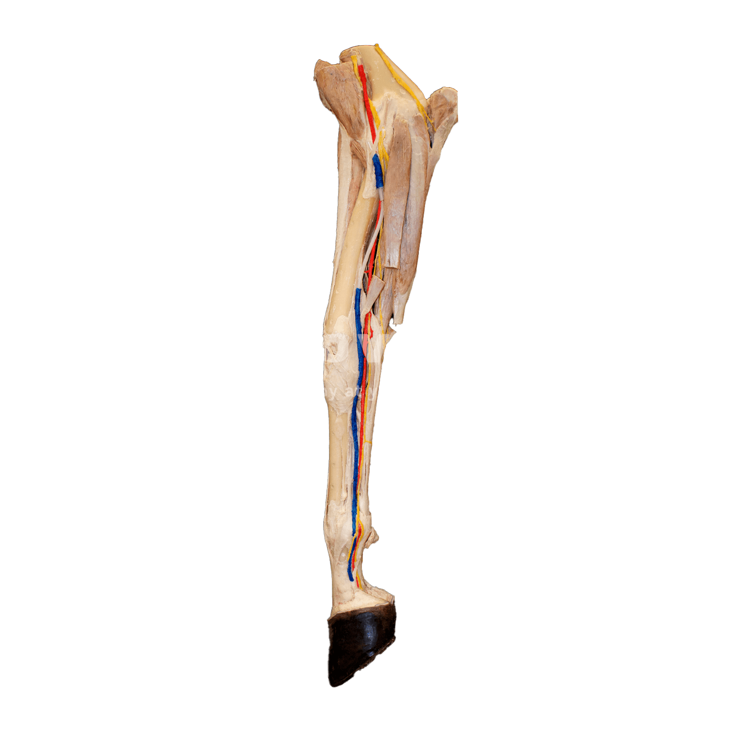
- Radius and Ulna: The radius bears most of the weight, while the ulna is partially fused to it in adult horses.
- Carpal bones (knee): Eight small bones arranged in three rows, allowing shock absorption during motion.
- Metacarpals: The third metacarpal (“cannon bone”) supports nearly 60–65% of the horse’s body weight, while the second and fourth (“splint bones”) stabilize the fetlock joint.
- Phalanges: Proximal, middle, and distal bones end in the hoof, which protects and cushions the distal limb.
This configuration gives the horse both stability and speed, but also makes it prone to degenerative and traumatic injuries if not properly managed.
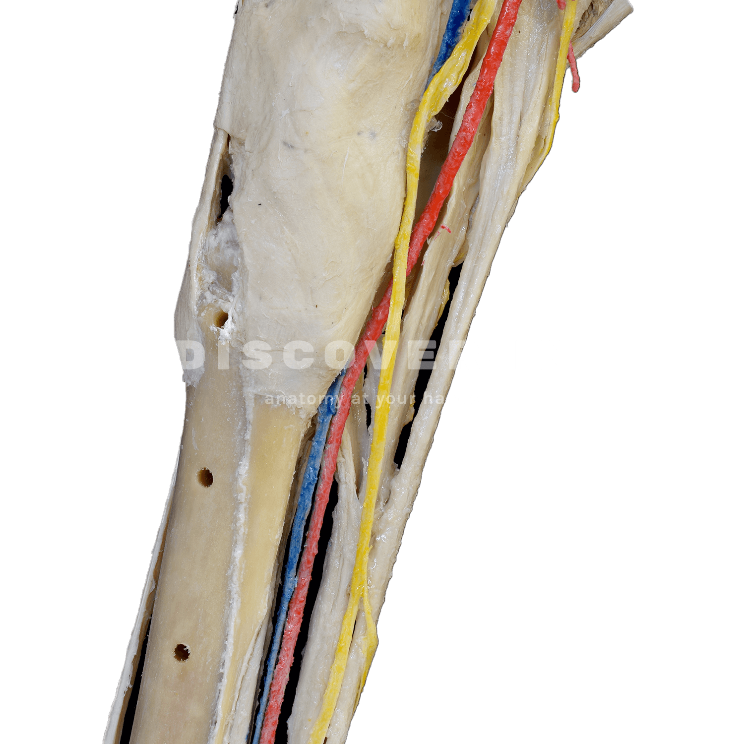
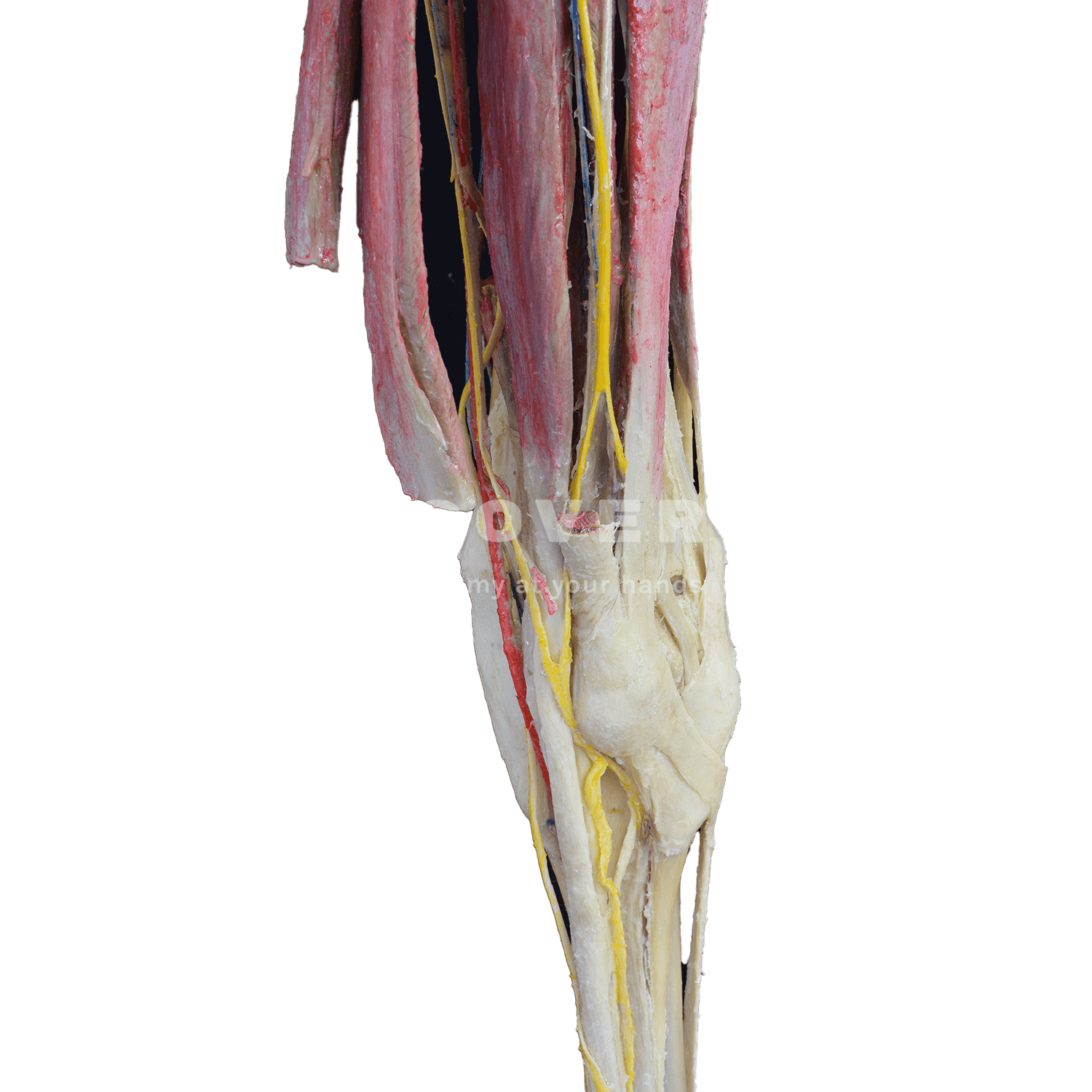
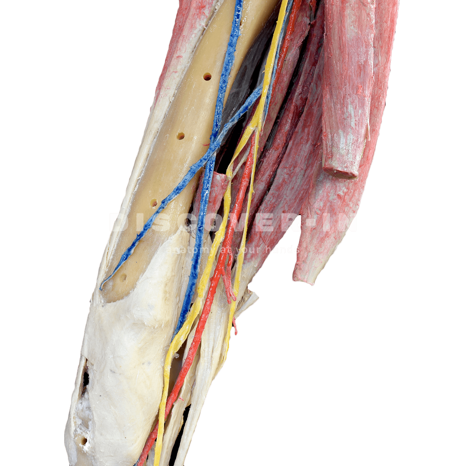
2. Muscles, Tendons, and Ligaments
The forelimb’s power and stability depend on proximal muscles and distal tendons that store and release energy during motion:
- Key muscles: Biceps brachii, triceps brachii, flexor and extensor groups of the carpus and digits.
- Critical tendons: Superficial and deep digital flexor tendons and the suspensory ligament—responsible for supporting the fetlock and preventing hyperextension.
Tendinitis and ligament injuries represent up to 40% of all musculoskeletal disorders in performance horses, highlighting the importance of proper anatomical understanding.
3. Functional Regions
Dividing the forelimb into functional regions enhances diagnosis and treatment planning:
- Shoulder: Muscle-heavy and prone to soft tissue injuries.
- Elbow: Occasionally affected by fractures or joint inflammation.
- Carpus (knee): Frequent site of osteoarthritis and chip fractures in racehorses.
- Metacarpus and Phalanges: Commonly involved in tendon, ligament, and hoof pathologies.
4. Nerves and Circulation
The brachial plexus gives rise to major nerves controlling motion and sensation:
- Radial nerve: Extends the carpus and digits.
- Median and ulnar nerves: Control flexion and palmar sensitivity.
- Cutaneous branches: Detect ground texture and prevent skin injuries.
Blood supply is dominated by the brachial artery, which divides into the radial and median arteries, continuing distally to digital arteries that help regulate hoof temperature and circulation.
5. Anatomical Terminology
Precise terminology ensures consistent communication between professionals:
- Forelimb / Front limb: Clinical reference.
- Medial / Lateral: Inner and outer surfaces.
- Proximal / Distal: Near or away from the body.
- Cranial / Caudal: Front and back surfaces.
6. Clinical and Practical Importance
Comprehensive forelimb anatomy knowledge is fundamental for:
- Lameness diagnosis: Pinpointing sources of pain and dysfunction.
- Surgical procedures: Understanding structural relationships ensures safety and precision.
- Rehabilitation: Designing targeted physiotherapy and exercise plans.
- Performance optimization: Healthy forelimbs are vital for speed, balance, and endurance.
Statistics reveal that over 80% of equine lameness cases originate in the forelimbs, often due to repetitive strain, hoof imbalance, or joint degeneration. Moreover, imaging studies show that early detection of microlesions can reduce recovery times by up to 50% when combined with preventive anatomical assessment.
The equine forelimb is a masterpiece of anatomical engineering — resilient yet vulnerable. A deep anatomical understanding not only supports accurate diagnosis and treatment but also enhances equine welfare and performance. Integrating plastinated anatomical models into veterinary education bridges the gap between theory and practice, allowing students and clinicians to visualize, handle, and study real structures safely and effectively, strengthening both academic learning and clinical expertise.