If you have access to our plastinated specimens, you will be able to deepen your anatomical knowledge in a practical and hands-on way.These specimens will enable you to:
• Precisely identify key anatomical structures.
• Understand anatomical reality in detail.
• Reinforce theoretical learning.
• Strengthen the acquisition of practical and clinical skills.
These advantages make Discover-IN specimens an innovative resource for students, educators, and professionals seeking comprehensive, effective, long-lasting, and biologically safe training in veterinary anatomy.
Discover-IN provides real plastinated specimens designed specifically for practical, safe, and long-lasting veterinary anatomy education and training. These materials are ideal for veterinary faculties, anatomy laboratories, and continuing education centers that require high-quality anatomical resources.
The catalog includes a wide range of species and anatomical systems, offering specimens that support hands-on understanding—from isolated organs to complex dissections, body sections or organ blocks. Each piece is unique thanks to the plastination process.
This resource adds significant value to veterinary training by enhancing learning through direct experience and complementing theoretical knowledge with real, easy-to-handle materials.
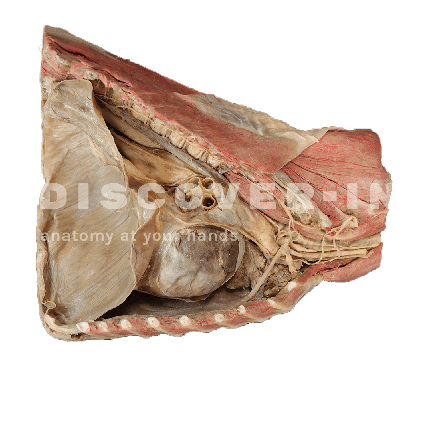
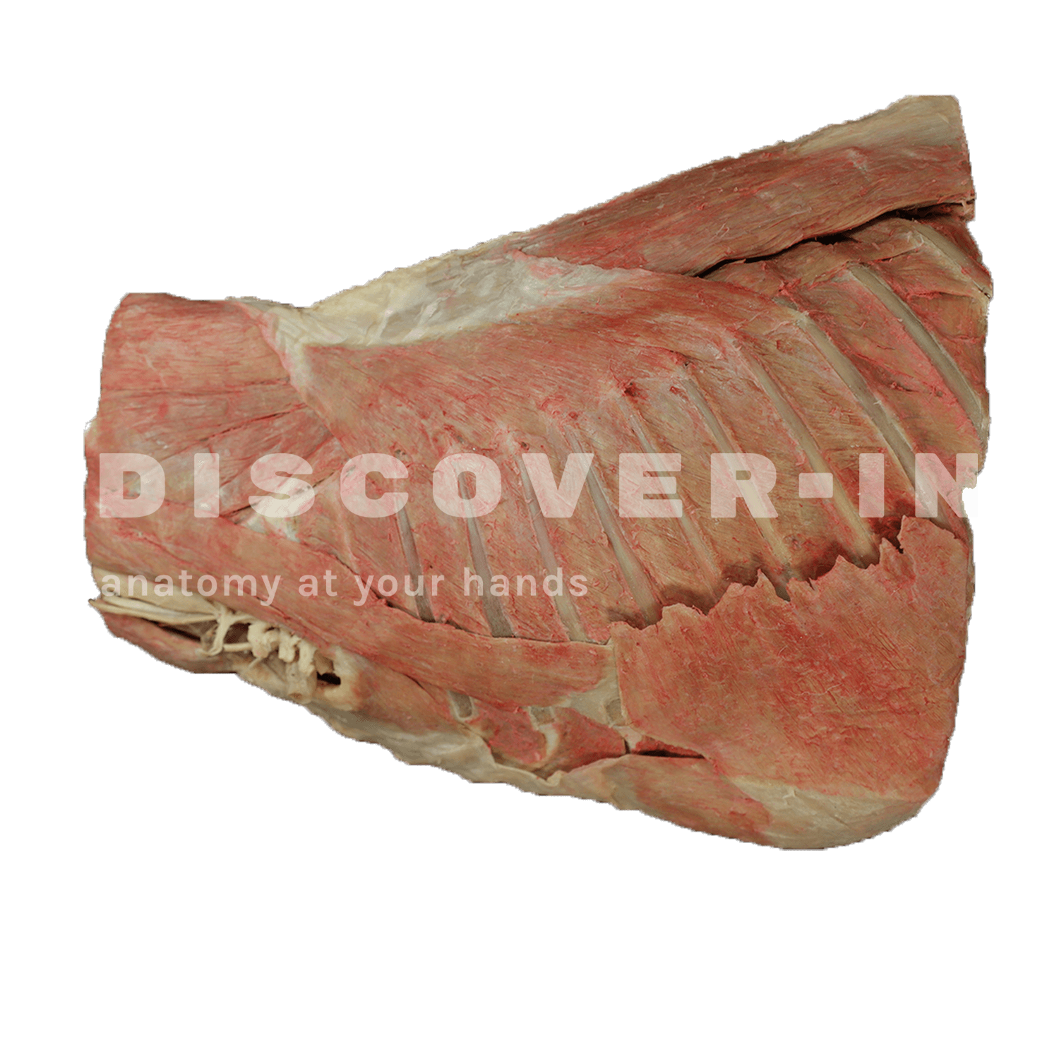
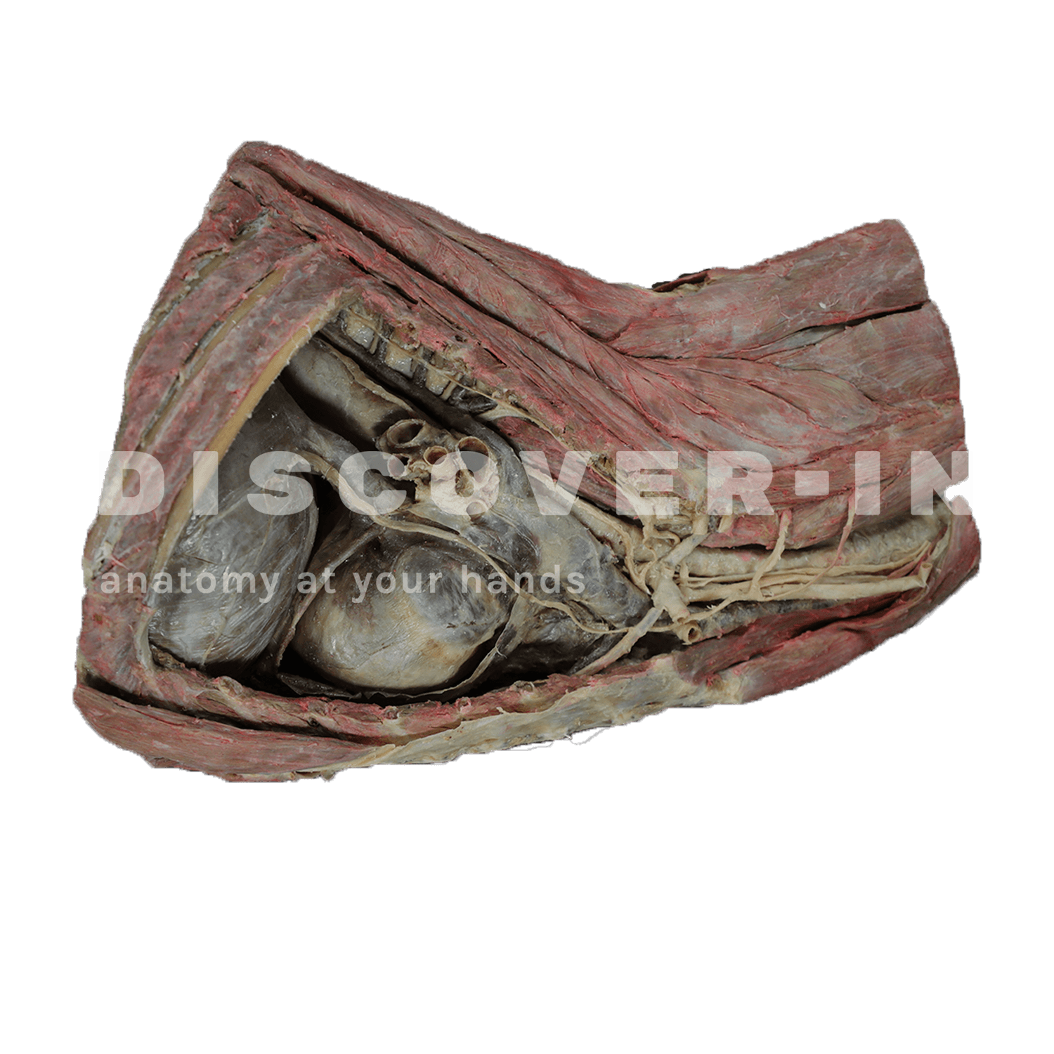
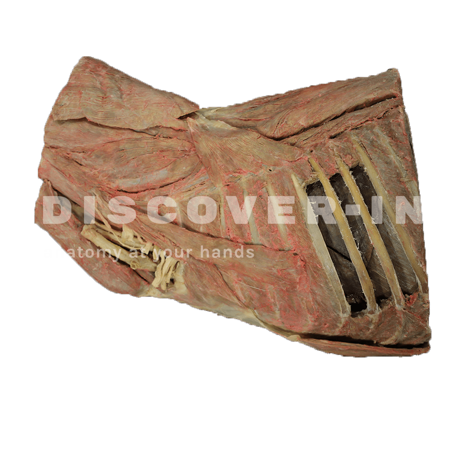
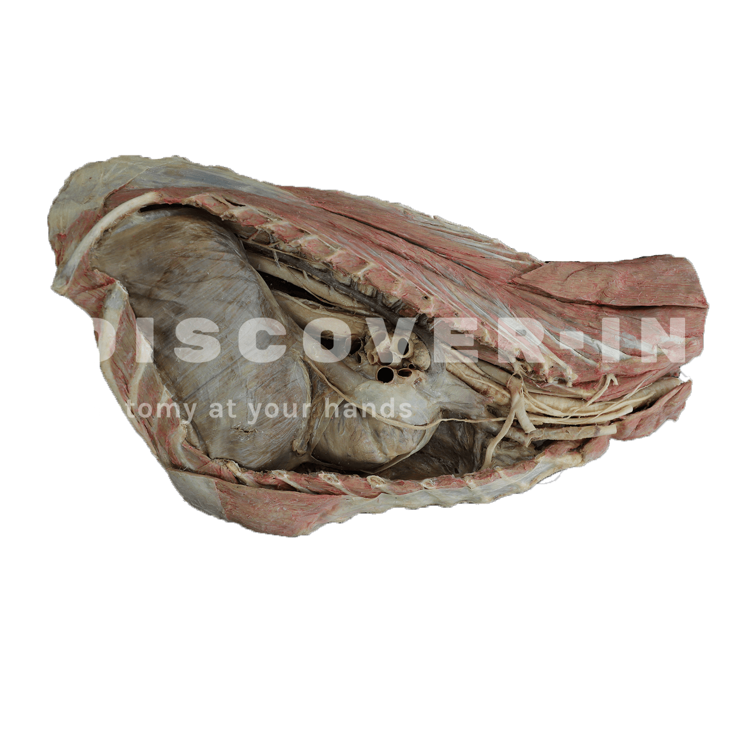
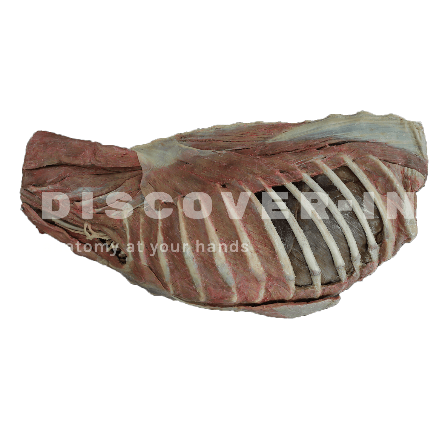
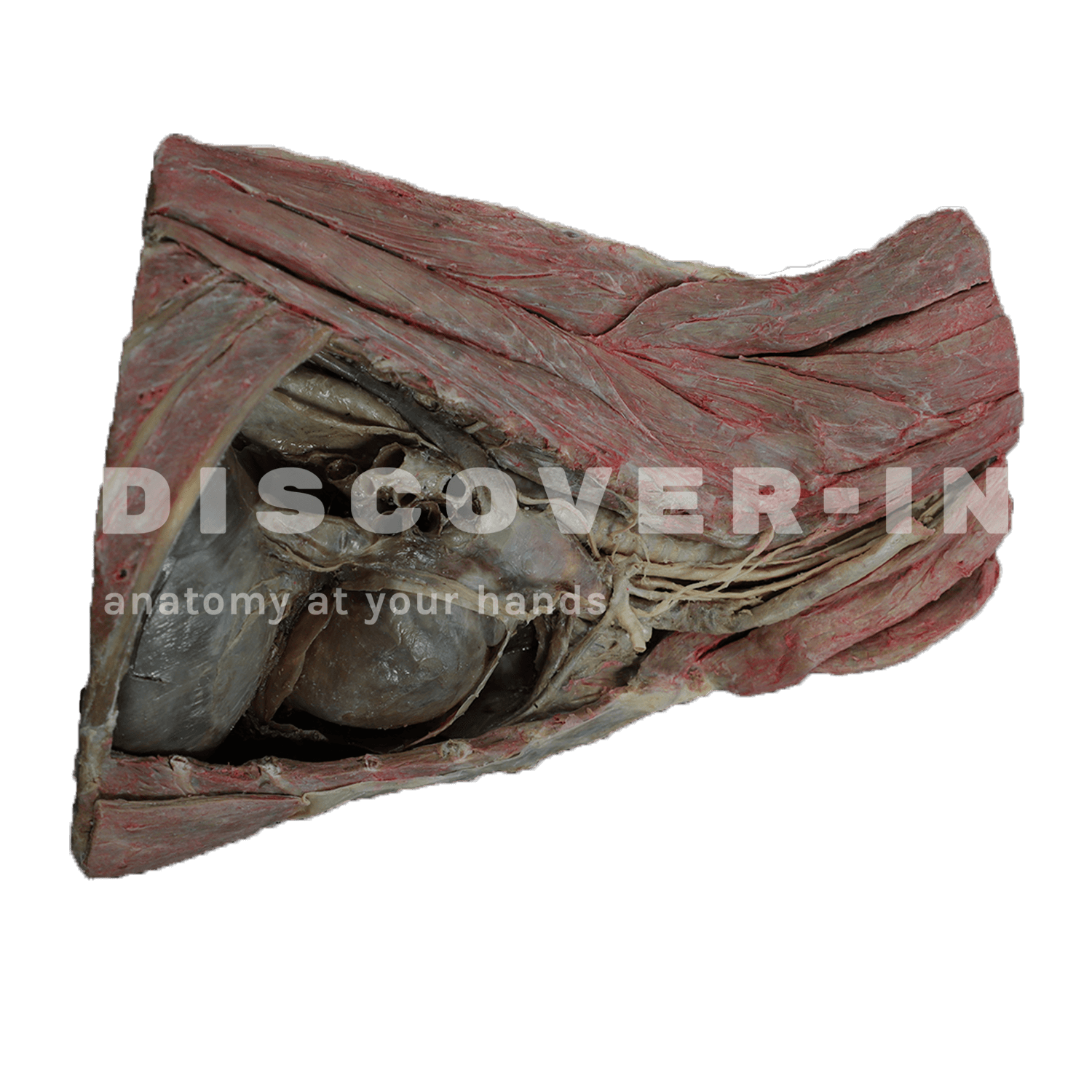
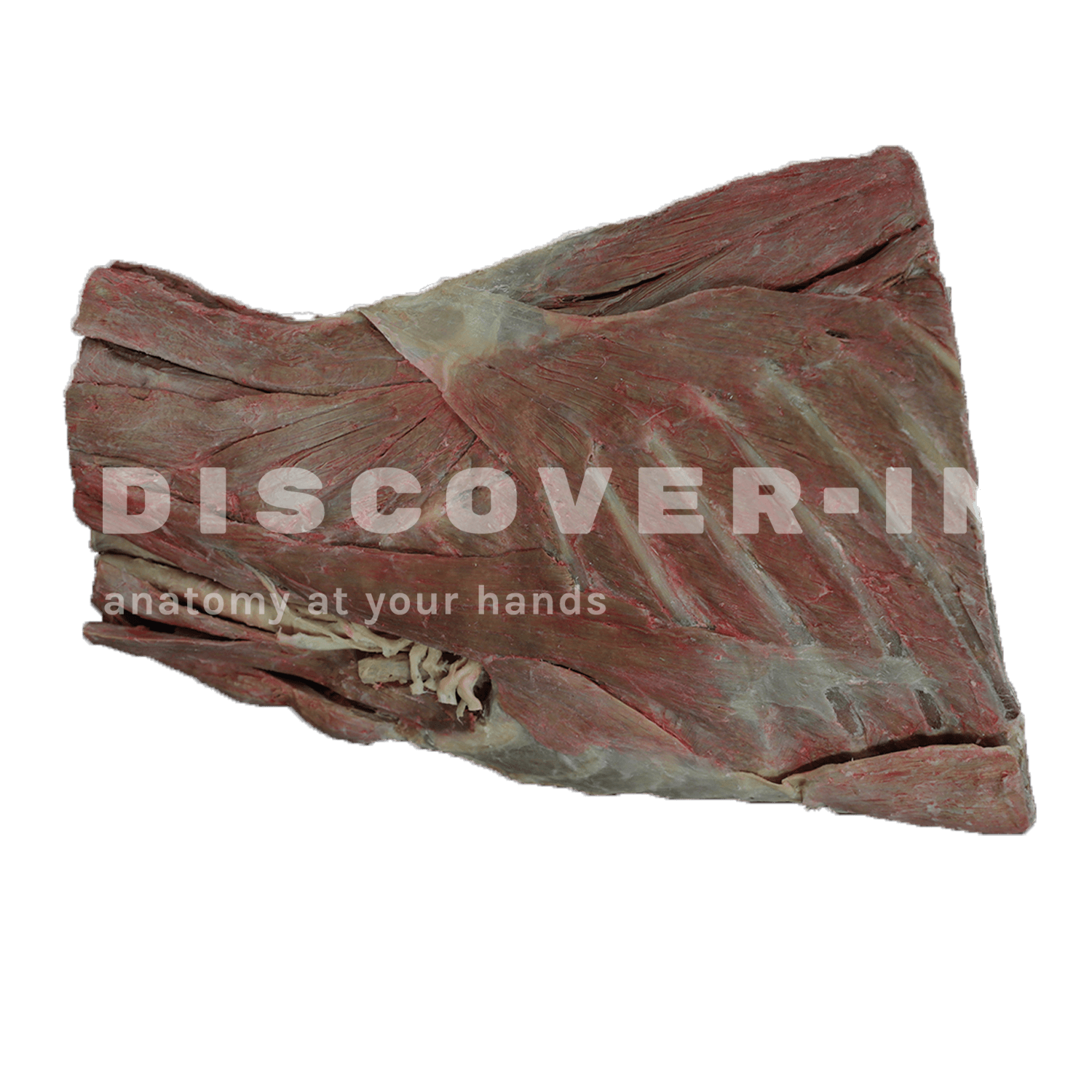
This dissection of the right thoracic cavity of a dog shows the mediastinum and its main structures, the heart with its pericardium, relevant vascular structures such as the thoracic aorta, the pulmonary trunk (sectioned), the venae cavae and right pulmonary veins (sectioned), the trachea (sectioned) and bronchi, the esophagus, the phrenic nerve, vagus nerve, sympathetic trunk, subclavian ansa, among others… as well as the diaphragm.
Furthermore, this specimen is also available in a digital 3D and augmented reality version at:https://discoveranatomy-in.com/app/landing