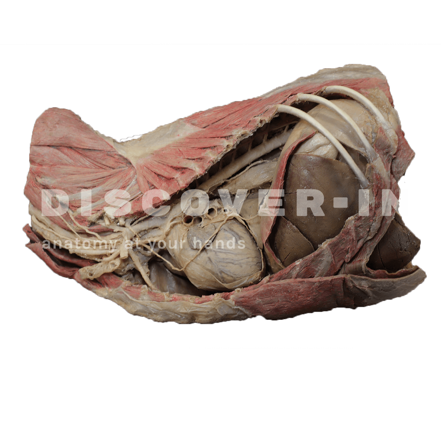
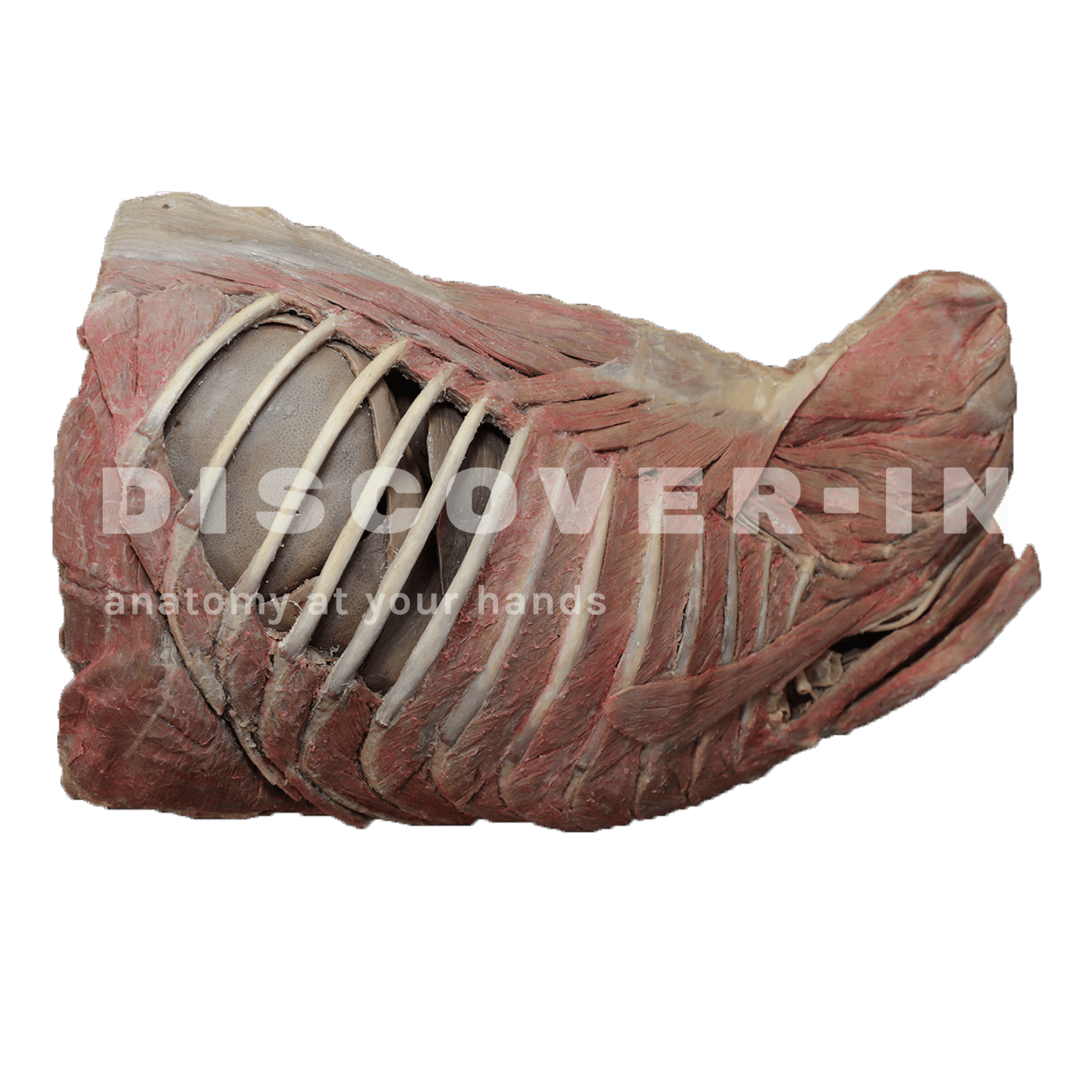
Canine thoracic cavity dissection left side and cranial abdomen
CaPtorIabdcr
More images
What is it in this specimen?
A left-sided plastinated dissection of the canine thoracic cavity with the thoracic wall opened to expose the pleural space and mediastinal contents. The ribs and intercostal musculature are visible along the opened thoracic wall, with the heart exposed in situ and superficial coronary (epicardial) vessels identifiable on the cardiac surface. Caudally, the thoracic cavity transitions to the cranial abdomen, where the diaphragm region is opened/removed sufficiently to allow visualization of cranial abdominal viscera—most consistently the liver (dark, lobated parenchyma) immediately caudal to the diaphragm.
What can we learn from this specimen?
- Topographic anatomy: appreciate the left lateral relationships between thoracic wall → pleural cavity → mediastinum (heart) and the caudal continuity with the diaphragm and cranial abdominal organs.
- Cardiac orientation: recognize the in situ position of the heart within the mediastinum and identify epicardial coronary vessels as clinically relevant landmarks on the cardiac surface.
- Thoracoabdominal boundary: understand how the diaphragm separates thoracic and abdominal cavities, and how cranial abdominal organs (notably the liver) lie immediately caudal to it.
How can this specimen be used for teaching?
- Clinical approach planning: discuss safe left intercostal access to the pleural cavity (thoracocentesis/chest drain concepts) by relating thoracic wall layers and intrathoracic organ proximity.
- Imaging correlation: correlate gross topography with thoracic radiographic/ultrasound expectations—heart position in the mediastinum and the cranial abdominal liver silhouette relative to the diaphragm.
- Surgical anatomy: use it to introduce principles of left thoracic exposure (thoracotomy orientation) and the practical importance of the diaphragm as the landmark for transitioning between thoracic and cranial abdominal surgical fields.