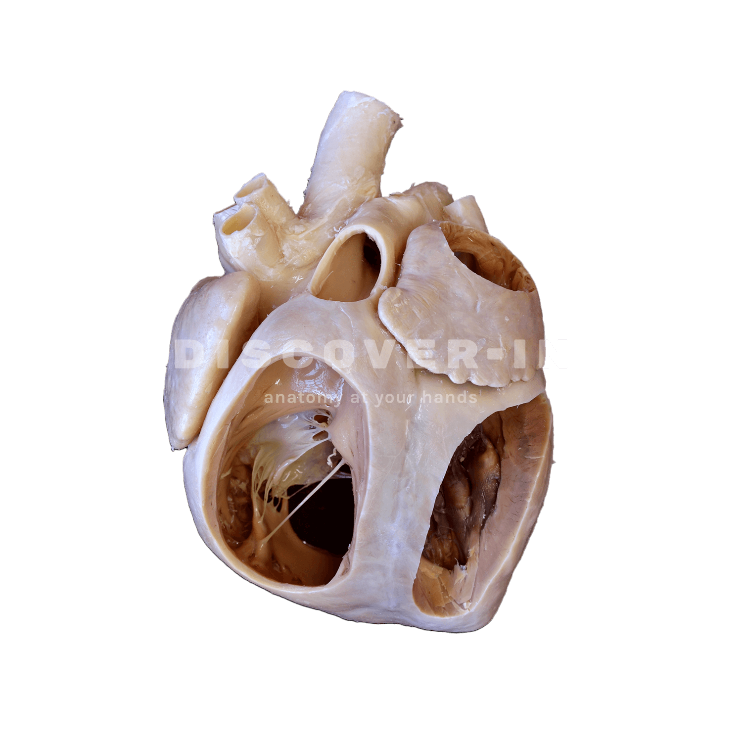
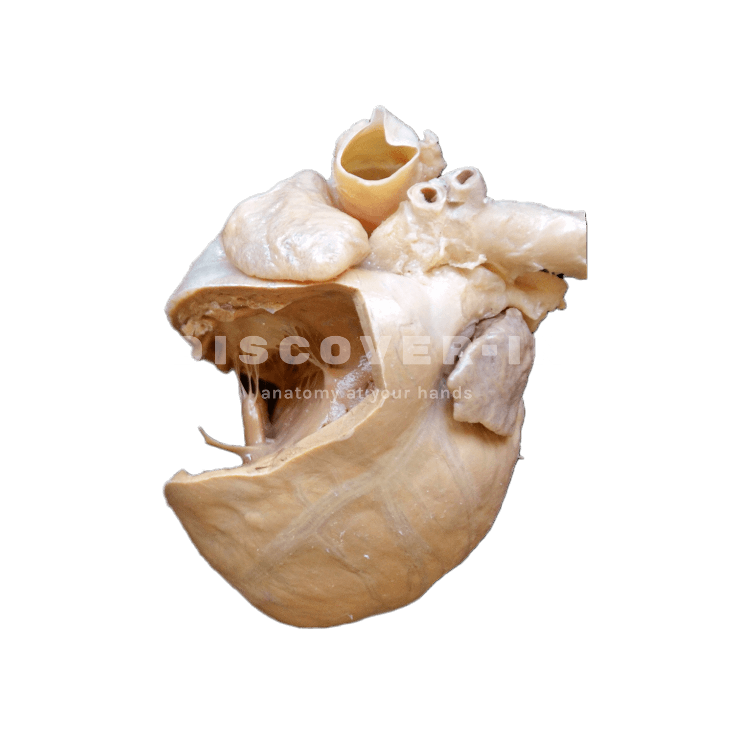
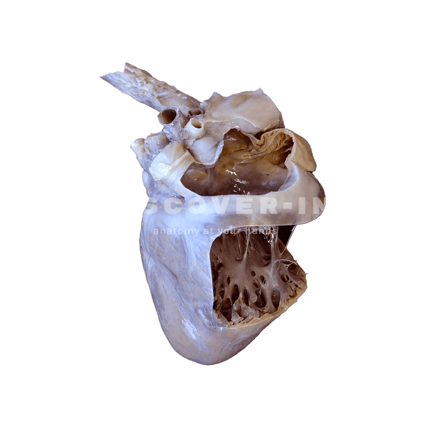
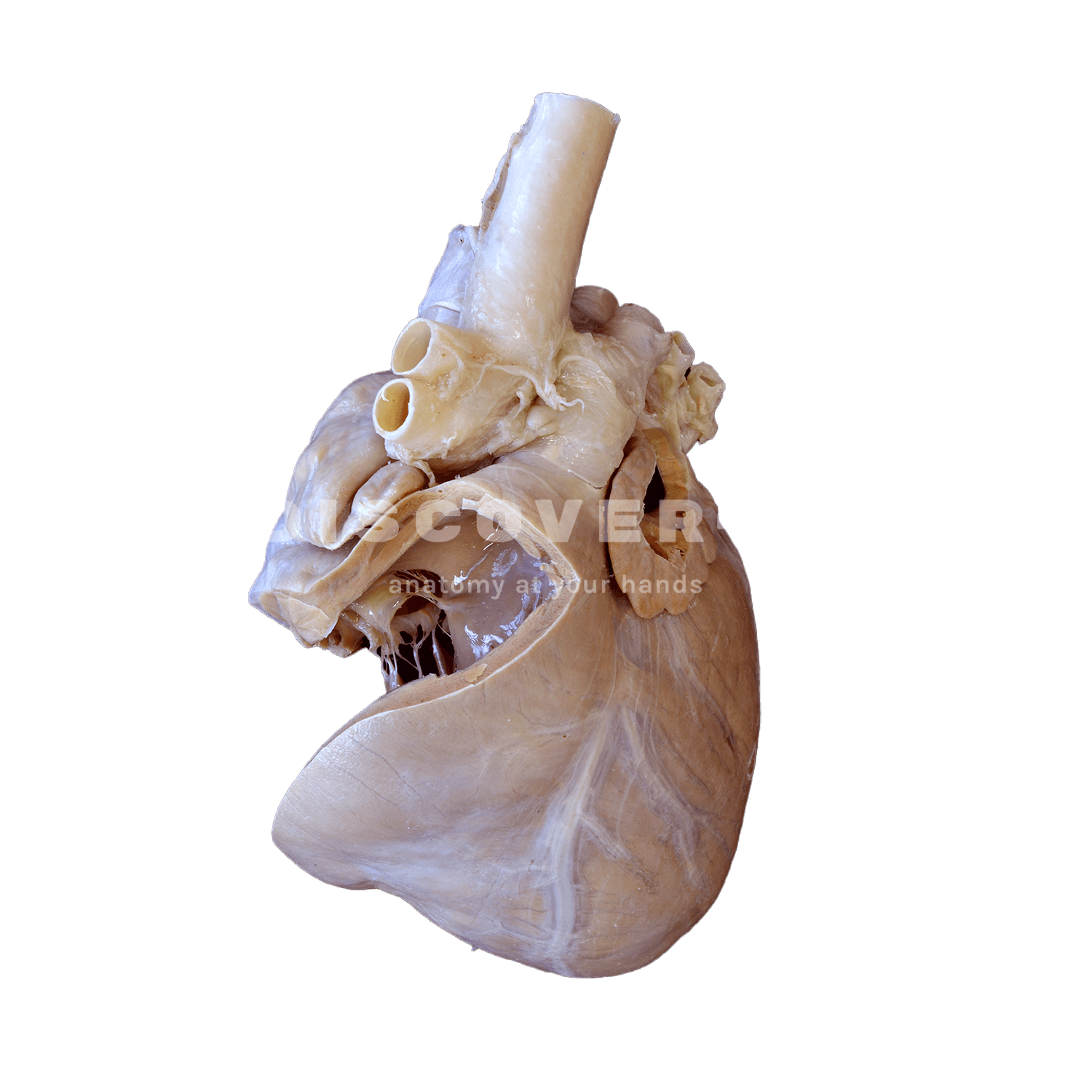
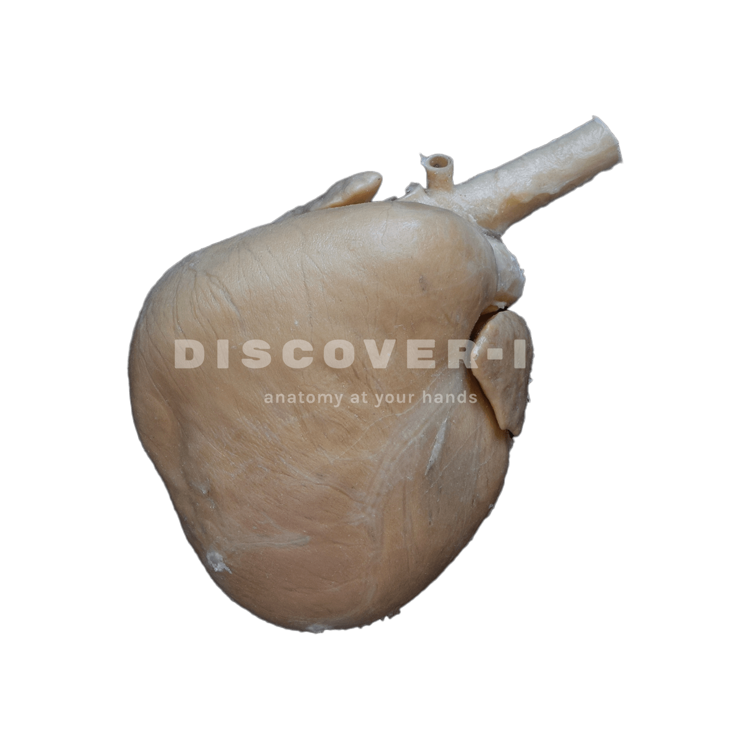
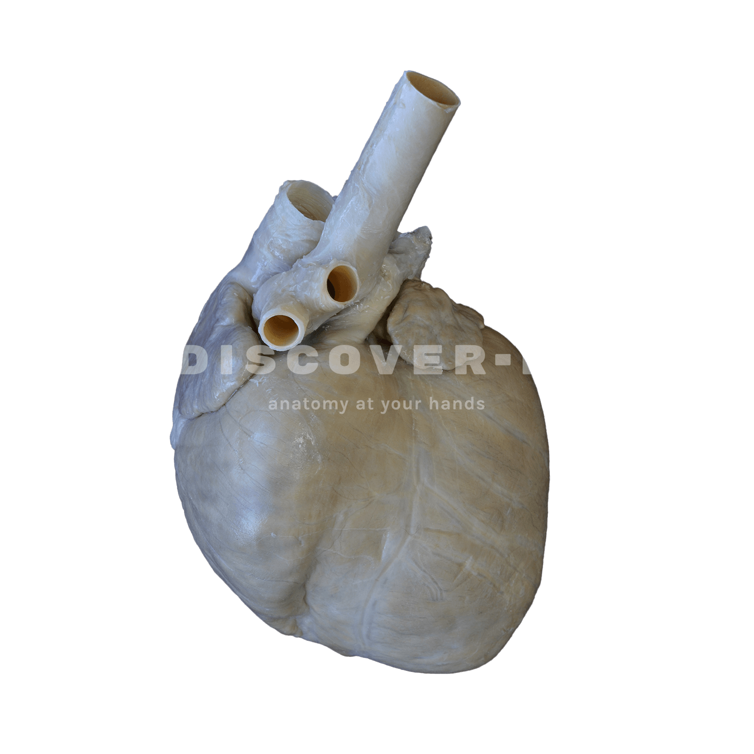
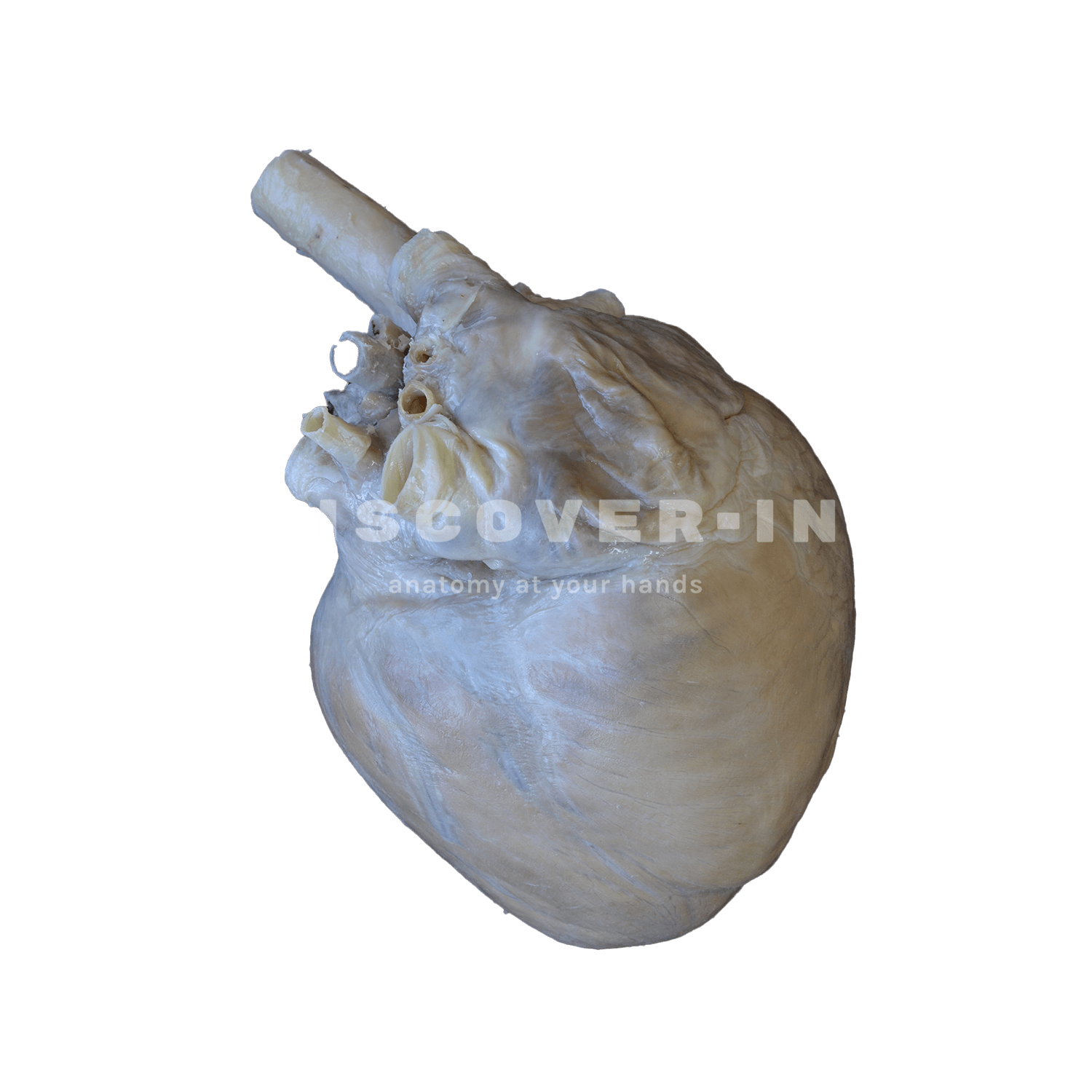
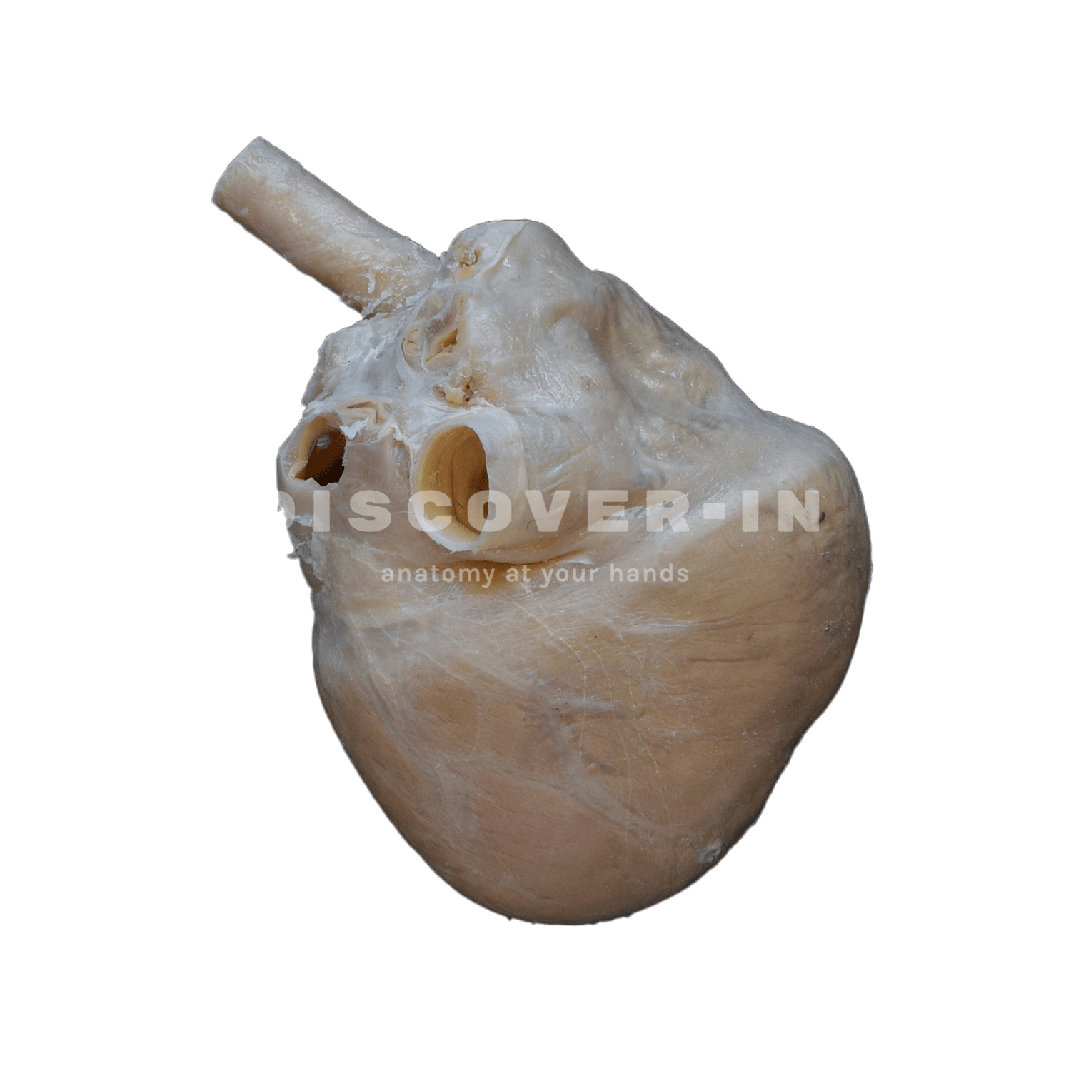
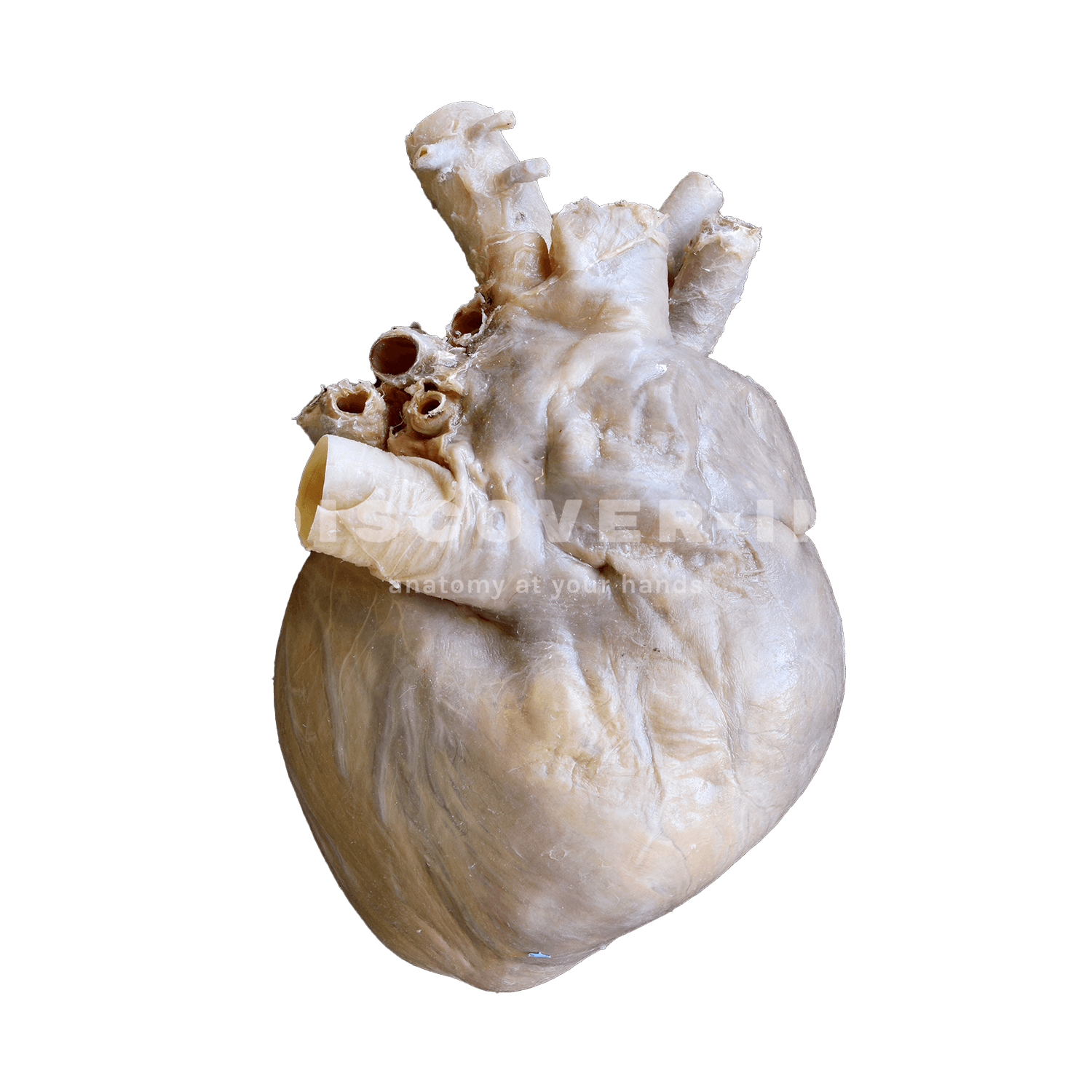
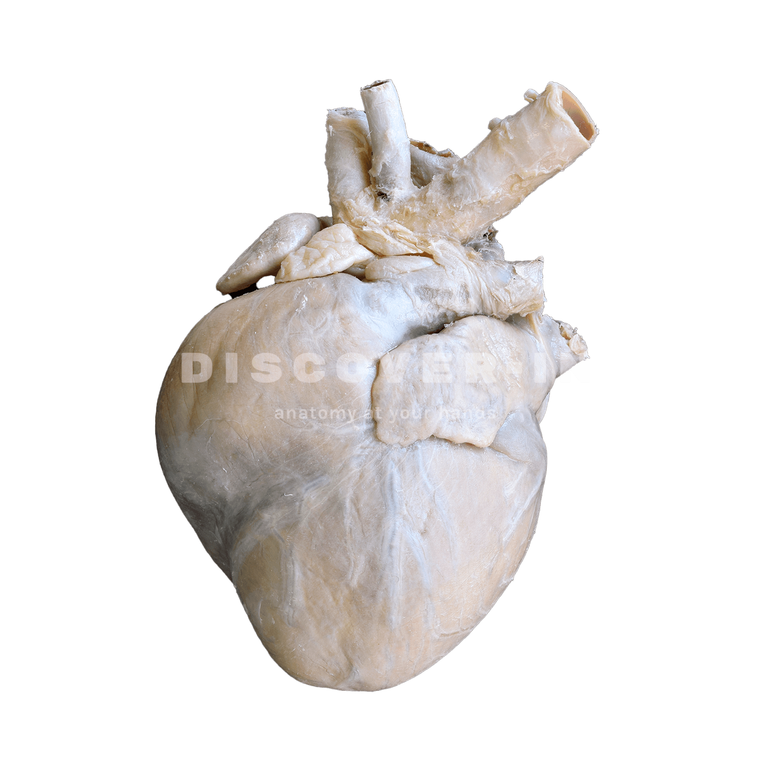
Plastinated canine heart showing heart chambers.
This specimen shows the anatomical characteristics of the external surface of the heart, including the atrial and atrial faces. At the base of the heart, the ascending aorta and its relationship with the pulmonary trunk are clearly observed, along with the entry points of the cranial and caudal caval veins to the right atrium. The bifurcation of the pulmonary trunk in the pulmonary arteries is observed, as well as the termination of the pulmonary veins in the left atrium. Ventricular and coronary grooves are visible. A portion of the right atrial wall has been removed to expose the right atrioventricular valve (tricuspid), including the tendon cords and papillary muscles.
In addition, the septomarginal trabecula ( moderating band ) is observed within the right ventricle, as well as its trajectory from the ventricle to the pulmonary trunk. The left atrial wall is also open to show its light.