Canine brain with arteries
CaOencAa
More images
What is it in this specimen?
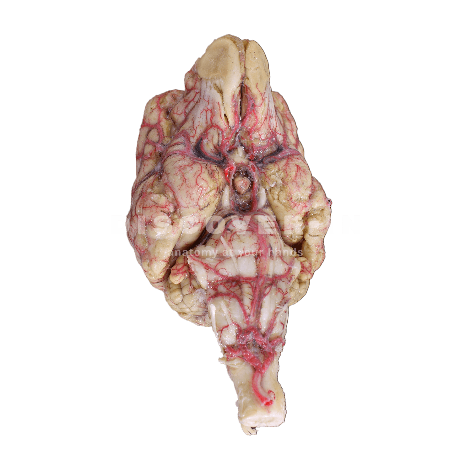
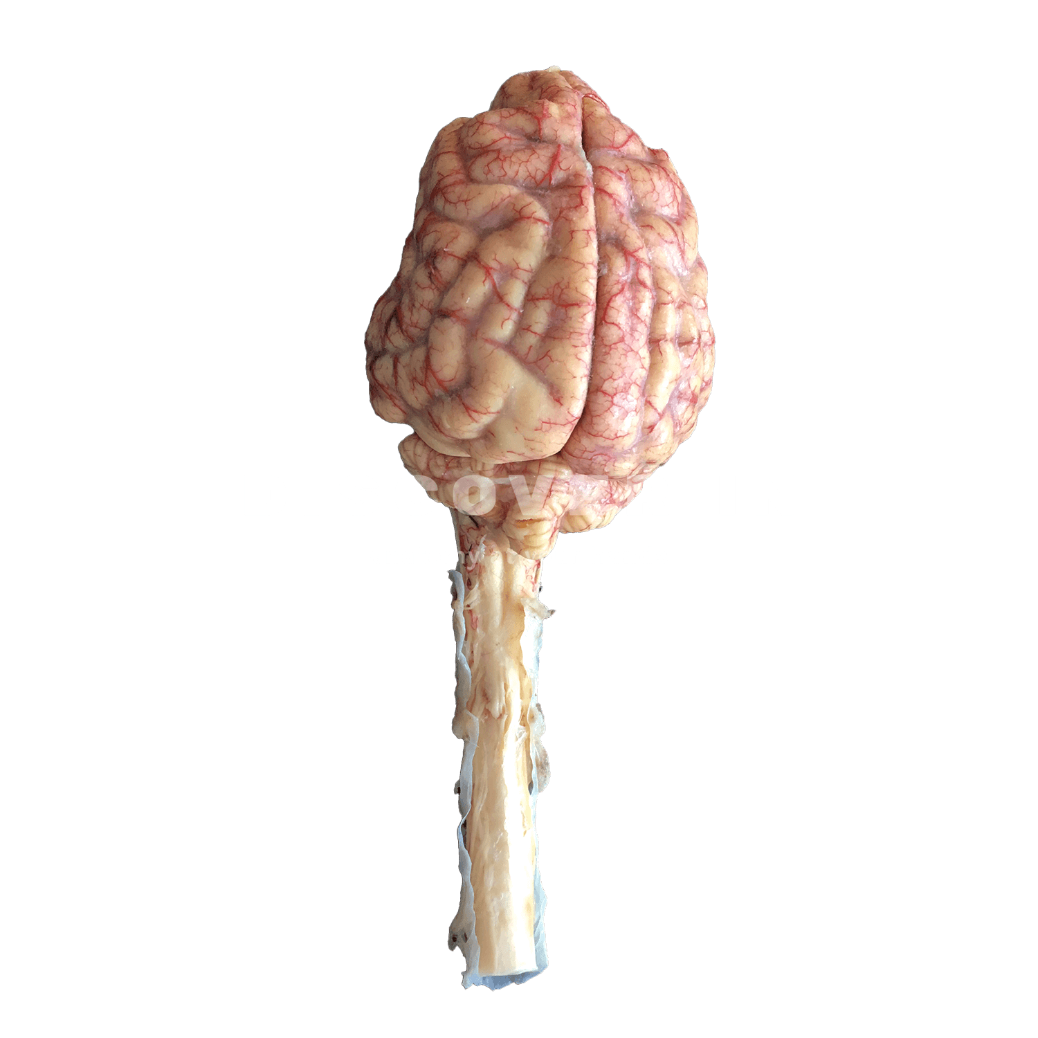
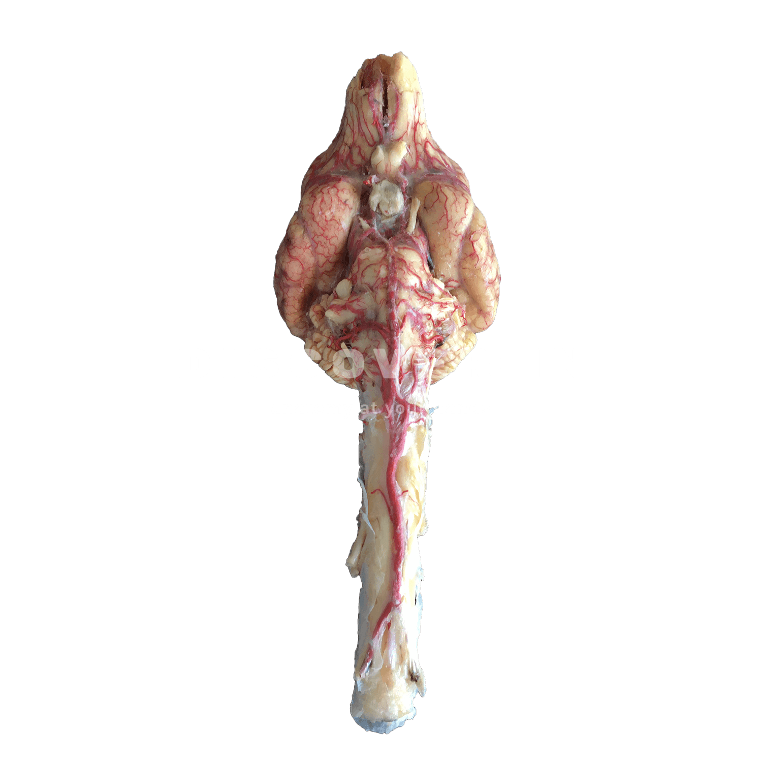
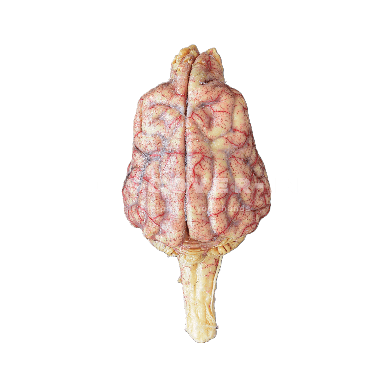
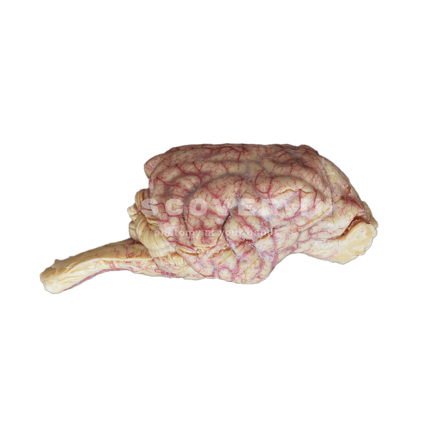
A plastinated canine brain with an injected/stained superficial arterial system, shown in dorsal and ventral aspects and including the brainstem. Fine leptomeningeal (pial) arteries are visible coursing over the cerebral hemispheres and across the cerebellar surface, while larger-calibre arteries are concentrated at the ventral base of the brain and along the ventral aspect of the brainstem, giving rise to branches that distribute rostrally over the cerebrum and caudally toward hindbrain territories.
What can we learn from this specimen?
- The canine cerebral circulation is organized with major arteries at the brain base and smaller pial branches spreading over the cortical and cerebellar surfaces.
- Vascular topography can be related to neuroanatomical regions (cerebral hemispheres vs cerebellum/brainstem), helping infer functionally relevant arterial “territories.”
- The dense packing of critical structures at the ventral brain base explains why lesions in this region can produce mixed cranial nerve and long-tract neurological signs.
How can this specimen be used for teaching?
- Correlate gross arterial distribution with neuroanatomical localization (forebrain signs vs brainstem/cerebellar signs) using the visible surface vessels as a guide.
- Support interpretation of CT/MRI by linking ventral brain landmarks (brain base and brainstem) to the course of major arteries and clinically important vascular regions.
- Discuss procedural and surgical risk in the ventral cranial cavity by highlighting why injury to basal arteries can rapidly compromise large portions of the CNS.